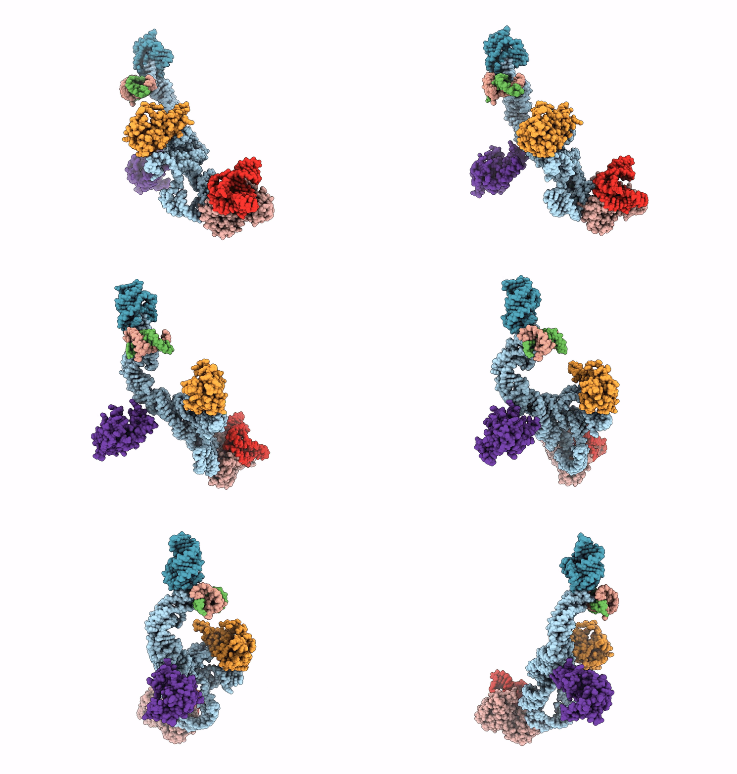
2NOQ image
Entry Detail
PDB ID:
2NOQ
Keywords:
Title:
Structure of ribosome-bound cricket paralysis virus IRES RNA
Biological Source:
Source Organism:
PDB Version:
Deposition Date:
2006-10-26
Release Date:
2006-11-21
Method Details:
Experimental Method:
Resolution:
7.30 Å
Aggregation State:
PARTICLE
Reconstruction Method:
SINGLE PARTICLE
Macromolecular Entities
Polymer Type:polyribonucleotide
Description:CrPV IRES
Chain IDs:A
Chain Length:190
Number of Molecules:1
Biological Source:
Polymer Type:polyribonucleotide
Description:18S ribosomal RNA
Chain IDs:B
Chain Length:46
Number of Molecules:1
Biological Source:Saccharomyces cerevisiae
Polymer Type:polyribonucleotide
Description:18S ribosomal RNA
Chain IDs:C
Chain Length:13
Number of Molecules:1
Biological Source:Saccharomyces cerevisiae
Polymer Type:polyribonucleotide
Description:18S ribosomal RNA
Chain IDs:D
Chain Length:15
Number of Molecules:1
Biological Source:Saccharomyces cerevisiae
Polymer Type:polyribonucleotide
Description:25S ribosomal RNA
Chain IDs:E
Chain Length:53
Number of Molecules:1
Biological Source:Saccharomyces cerevisiae
Polymer Type:polypeptide(L)
Description:40S ribosomal protein S5
Chain IDs:F
Chain Length:150
Number of Molecules:1
Biological Source:Saccharomyces cerevisiae
Polymer Type:polypeptide(L)
Description:60S ribosomal protein L1
Chain IDs:G
Chain Length:213
Number of Molecules:1
Biological Source:Saccharomyces cerevisiae
Polymer Type:polypeptide(L)
Description:60S ribosomal protein L11-B
Chain IDs:H
Chain Length:165
Number of Molecules:1
Biological Source:Saccharomyces cerevisiae
Ligand Molecules
Primary Citation
Structure of the ribosome-bound cricket paralysis virus IRES RNA.
Nat.Struct.Mol.Biol. 13 1092 1096 (2006)
PMID: 17115051 DOI: 10.1038/nsmb1177

Abstact

Internal ribosome entry sites (IRESs) facilitate an alternative, end-independent pathway of translation initiation. A particular family of dicistroviral IRESs can assemble elongation-competent 80S ribosomal complexes in the absence of canonical initiation factors and initiator transfer RNA. We present here a cryo-EM reconstruction of a dicistroviral IRES bound to the 80S ribosome. The resolution of the cryo-EM reconstruction, in the subnanometer range, allowed the molecular structure of the complete IRES in its active, ribosome-bound state to be solved. The structure, harboring three pseudoknot-containing domains, each with a specific functional role, shows how defined elements of the IRES emerge from a compactly folded core and interact with the key ribosomal components that form the A, P and E sites, where tRNAs normally bind. Our results exemplify the molecular strategy for recruitment of an IRES and reveal the dynamic features necessary for internal initiation.

Legend

Protein

Chemical

Disease

Primary Citation of related structures