Deposition Date
2006-10-23
Release Date
2007-01-16
Last Version Date
2023-10-25
Entry Detail
PDB ID:
2NMV
Keywords:
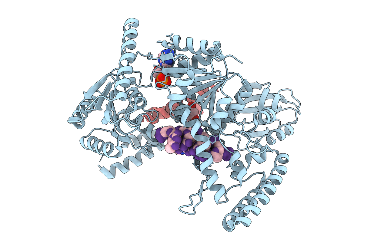
Title:
Damage detection by the UvrABC pathway: Crystal structure of UvrB bound to fluorescein-adducted DNA
Biological Source:
Source Organism(s):
Bacillus subtilis (Taxon ID: 1423)
Expression System(s):
Method Details:
Experimental Method:
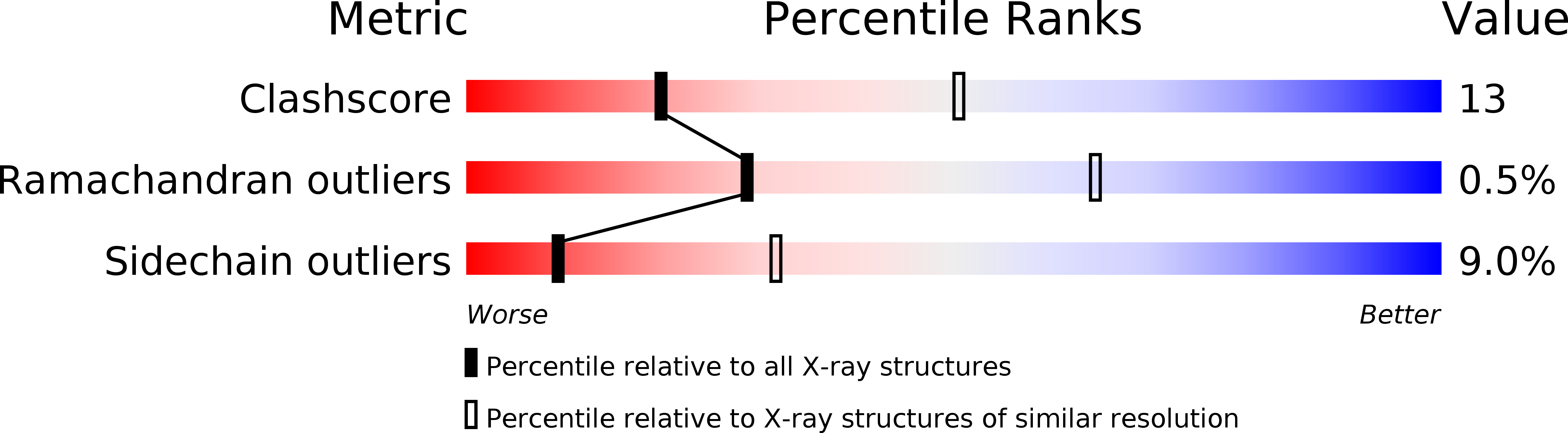
Resolution:
2.95 Å
R-Value Free:
0.28
R-Value Work:
0.22
R-Value Observed:
0.22
Space Group:
P 21 21 21