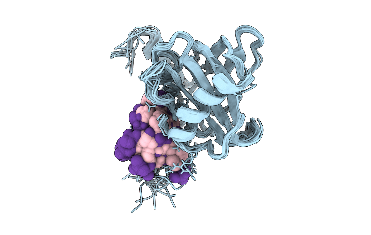
2NMB image
Deposition Date 1998-10-29
Release Date 1998-11-04
Last Version Date 2024-10-16
Entry Detail
PDB ID:
2NMB
Title:
DNUMB PTB DOMAIN COMPLEXED WITH A PHOSPHOTYROSINE PEPTIDE, NMR, ENSEMBLE OF STRUCTURES.
Biological Source:
Source Organism:
Method Details:
Experimental Method:
Conformers Calculated:
200
Conformers Submitted:
14
Selection Criteria:
NO NOE VIOLATION > 0.3 A
Macromolecular Entities
Polymer Type:polypeptide(L)
Molecule:PROTEIN (NUMB PROTEIN)
Gene (Uniprot):numb
Chain IDs:A
Chain Length:160
Number of Molecules:1
Biological Source:Drosophila melanogaster
Modified Residue
Compound ID Chain ID Parent Comp ID Details 2D Image
PTR B TYR O-PHOSPHOTYROSINE
Ligand Molecules
Primary Citation
Structure of a Numb PTB domain-peptide complex suggests a basis for diverse binding specificity.
Nat.Struct.Biol. 5 1075 1083 (1998)
PMID: 9846878 DOI: 10.1038/4185

Abstact

The phosphotyrosine-binding (PTB) domain of Numb, a protein involved in asymmetric cell division, has recently been shown to bind to the adapter protein Lnx through an LDNPAY sequence, to the Numb-associated kinase (Nak) through a sequence that does not contain an NPXY motif and to GP(p)Y-containing peptides obtained from library screening. We show here that these diverse peptide sequences bind with comparable affinities to the Numb PTB domain at a common binding site on the surface of the protein. The NMR structure of the Numb PTB domain in complex with a GPpY-containing peptide reveals a novel mechanism of binding with the peptide in a helical turn that does not hydrogen bond to the PTB domain beta-sheet. These results suggest that PTB domains can potentially have multiple modes of peptide recognition and provide a structural basis from which the multiple functions of the Numb PTB domain during asymmetric cell division could arise.

Legend

Protein

Chemical

Disease

Primary Citation of related structures