2NM1 image
Deposition Date 2006-10-20
Release Date 2006-12-19
Last Version Date 2023-08-30
Entry Detail
PDB ID:
2NM1
Keywords:
Title:
Structure of BoNT/B in complex with its protein receptor
Biological Source:
Source Organism(s):
Expression System(s):
Method Details:
Experimental Method:
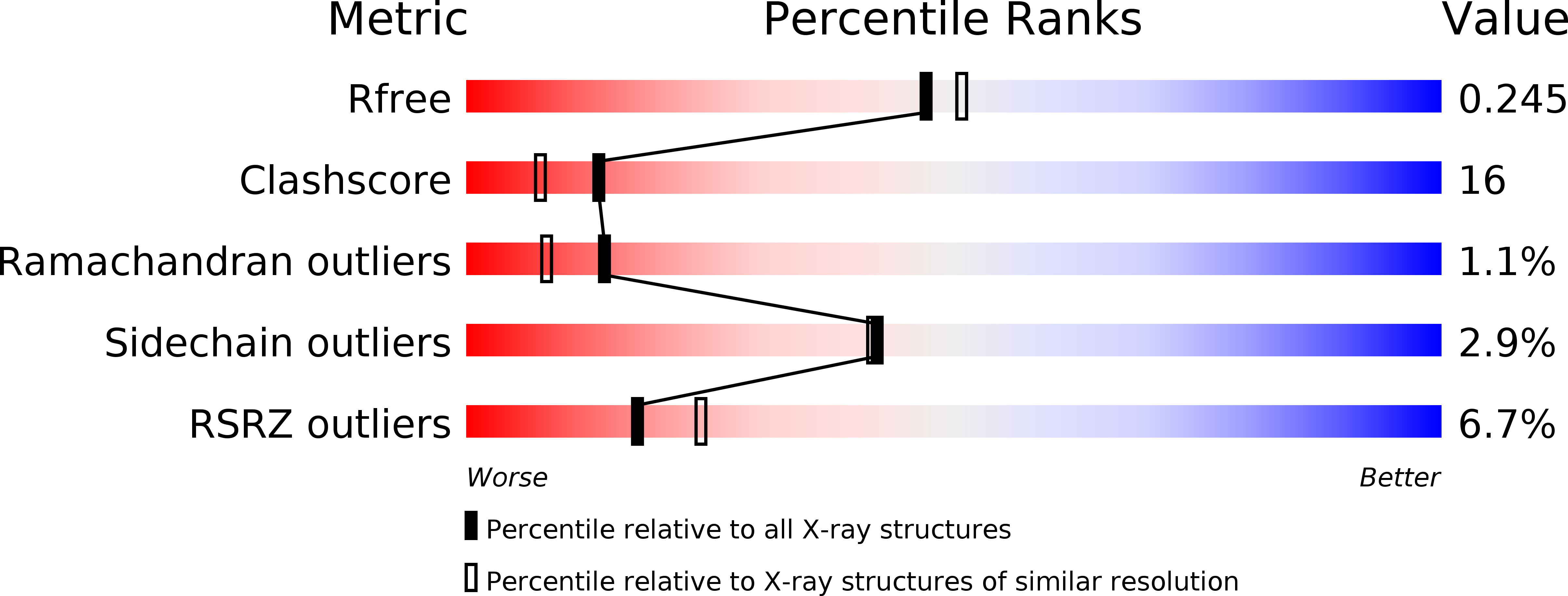
Resolution:
2.15 Å
R-Value Free:
0.24
R-Value Work:
0.19
R-Value Observed:
0.19
Space Group:
P 21 21 21
Macromolecular Entities
Structures with similar UniProt ID
Protein Blast
Polymer Type:polypeptide(L)
Molecule:Botulinum neurotoxin type B
Gene (Uniprot):botB
Chain IDs:A
Chain Length:436
Number of Molecules:1
Biological Source:Clostridium botulinum
Structures with similar UniProt ID
Protein Blast
Polymer Type:polypeptide(L)
Molecule:Synaptotagmin-2
Chain IDs:B
Chain Length:17
Number of Molecules:1
Biological Source:Rattus norvegicus
Primary Citation
Botulinum neurotoxin B recognizes its protein receptor with high affinity and specificity.
Nature 444 1092 1095 (2006)
PMID: 17167421 DOI: 10.1038/nature05387

Abstact

Botulinum neurotoxins (BoNTs) are produced by Clostridium botulinum and cause the neuroparalytic syndrome of botulism. With a lethal dose of 1 ng kg(-1), they pose a biological hazard to humans and a serious potential bioweapon threat. BoNTs bind with high specificity at neuromuscular junctions and they impair exocytosis of synaptic vesicles containing acetylcholine through specific proteolysis of SNAREs (soluble N-ethylmaleimide-sensitive fusion protein attachment protein receptors), which constitute part of the synaptic vesicle fusion machinery. The molecular details of the toxin-cell recognition have been elusive. Here we report the structure of a BoNT in complex with its protein receptor: the receptor-binding domain of botulinum neurotoxin serotype B (BoNT/B) bound to the luminal domain of synaptotagmin II, determined at 2.15 A resolution. On binding, a helix is induced in the luminal domain which binds to a saddle-shaped crevice on a distal tip of BoNT/B. This crevice is adjacent to the non-overlapping ganglioside-binding site of BoNT/B. Synaptotagmin II interacts with BoNT/B with nanomolar affinity, at both neutral and acidic endosomal pH. Biochemical and neuronal ex vivo studies of structure-based mutations indicate high specificity and affinity of the interaction, and high selectivity of BoNT/B among synaptotagmin I and II isoforms. Synergistic binding of both synaptotagmin and ganglioside imposes geometric restrictions on the initiation of BoNT/B translocation after endocytosis. Our results provide the basis for the rational development of preventive vaccines or inhibitors against these neurotoxins.

Legend

Protein

Chemical

Disease

Primary Citation of related structures
Feedback Form
Name
Email
Institute
Feedback