2NLL image
Deposition Date 1996-11-20
Release Date 1997-03-12
Last Version Date 2024-02-21
Entry Detail
PDB ID:
2NLL
Title:
RETINOID X RECEPTOR-THYROID HORMONE RECEPTOR DNA-BINDING DOMAIN HETERODIMER BOUND TO THYROID RESPONSE ELEMENT DNA
Biological Source:
Source Organism:
Homo sapiens (Taxon ID: 9606)
Host Organism:
Method Details:
Experimental Method:
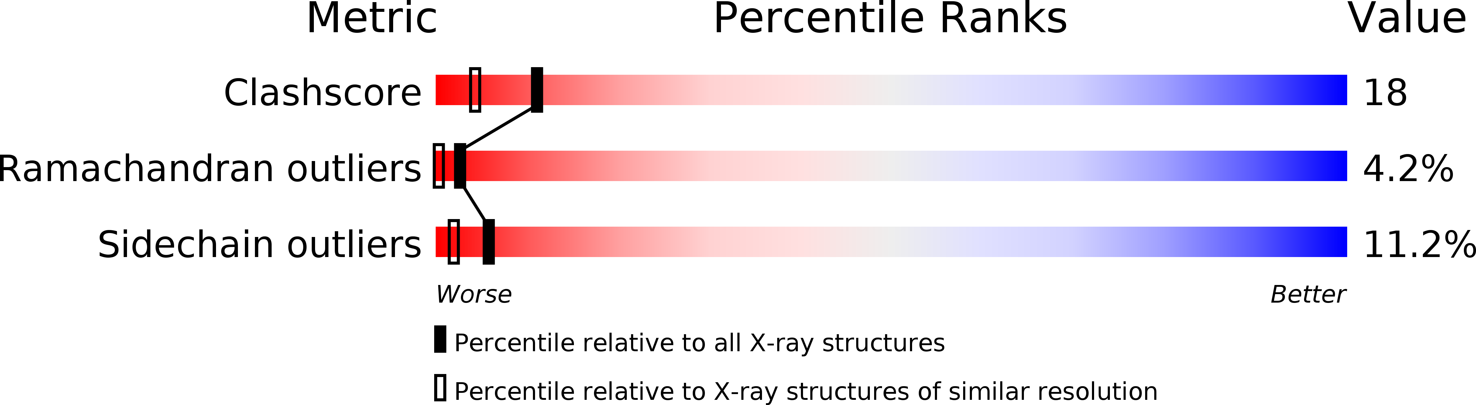
Resolution:
1.90 Å
R-Value Free:
0.26
R-Value Work:
0.20
R-Value Observed:
0.20
Space Group:
P 21 21 21
Macromolecular Entities
Structures with similar UniProt ID
Protein Blast
Polymer Type:polypeptide(L)
Molecule:PROTEIN (RETINOIC ACID RECEPTOR)
Gene (Uniprot):RXRA
Chain IDs:C (auth: A)
Chain Length:66
Number of Molecules:1
Biological Source:Homo sapiens
Structures with similar UniProt ID
Protein Blast
Polymer Type:polypeptide(L)
Molecule:PROTEIN (THYROID HORMONE RECEPTOR)
Gene (Uniprot):THRB
Chain IDs:D (auth: B)
Chain Length:103
Number of Molecules:1
Biological Source:Homo sapiens
Modified Residue
Compound ID Chain ID Parent Comp ID Details 2D Image
5IU A DU 5-IODO-2'-DEOXYURIDINE-5'-MONOPHOSPHATE
Ligand Molecules
Primary Citation
Structural determinants of nuclear receptor assembly on DNA direct repeats.
Nature 375 203 211 (1995)
PMID: 7746322 DOI: 10.1038/375203a0

Abstact

Nuclear receptor heterodimers recognize response elements composed of two direct repeats of the consensus sequence 5'-AGGTCA-3' separated by one to five base pairs. The 1.9 A crystal structure of the complex formed by the DNA-binding domains of the 9-cis retinoic acid receptor and thyroid hormone receptor bound to a thyroid-response element shows that the subunits interact through a DNA-supported interface involving the carboxy-terminal extension of the DNA-binding domain of the thyroid hormone receptor. The stereochemistry suggests a mechanism by which heterodimers recognize the inter-half-site spacing between direct repeats.

Legend

Protein

Chemical

Disease

Primary Citation of related structures
Feedback Form
Name
Email
Institute
Feedback