Deposition Date
2015-09-06
Release Date
2015-10-28
Last Version Date
2024-05-15
Entry Detail
PDB ID:
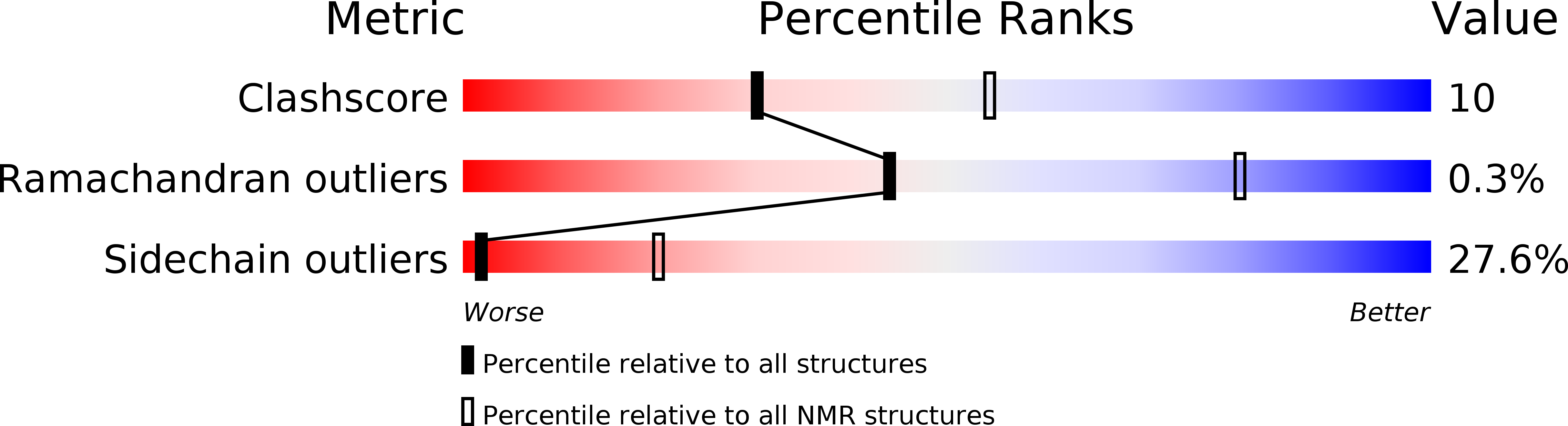
2N79
Keywords:
Title:
The structural and functional effects of the Familial Hypertrophic Cardiomyopathy-linked cardiac troponin C mutation, L29Q
Biological Source:
Source Organism(s):
Homo sapiens (Taxon ID: 9606)
Expression System(s):
Method Details:
Experimental Method:
Conformers Calculated:
100
Conformers Submitted:
20
Selection Criteria:
structures with the lowest energy