
Deposition Date
2015-08-07
Release Date
2015-11-25
Last Version Date
2024-11-20
Entry Detail
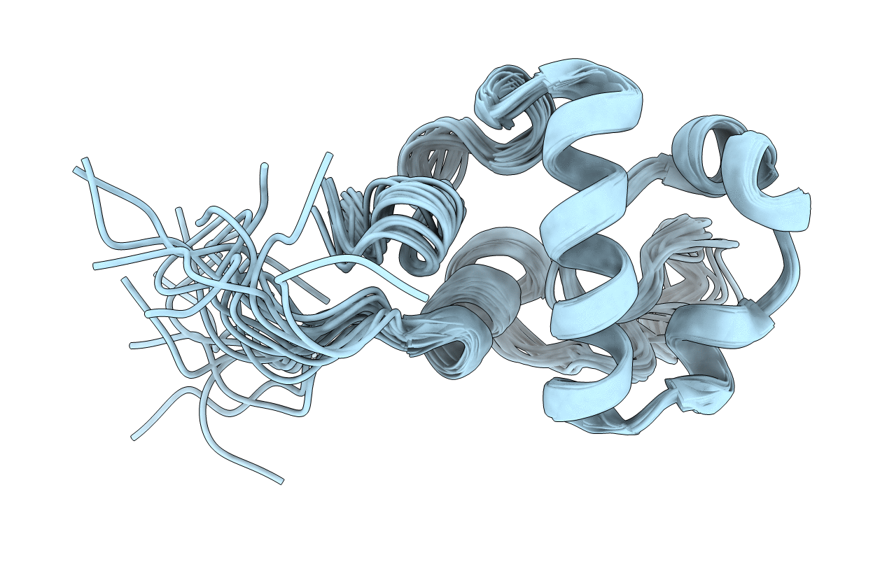
PDB ID:
2N5Z
Keywords:
Title:
Mycobacterium tuberculosis: a dynamic view of the resuscitation promoting factor RpfC catalytic domain
Biological Source:
Source Organism(s):
Mycobacterium tuberculosis (Taxon ID: 83332)
Expression System(s):
Method Details:
Experimental Method:
Conformers Calculated:
100
Conformers Submitted:
20
Selection Criteria:
structures with the lowest energy
