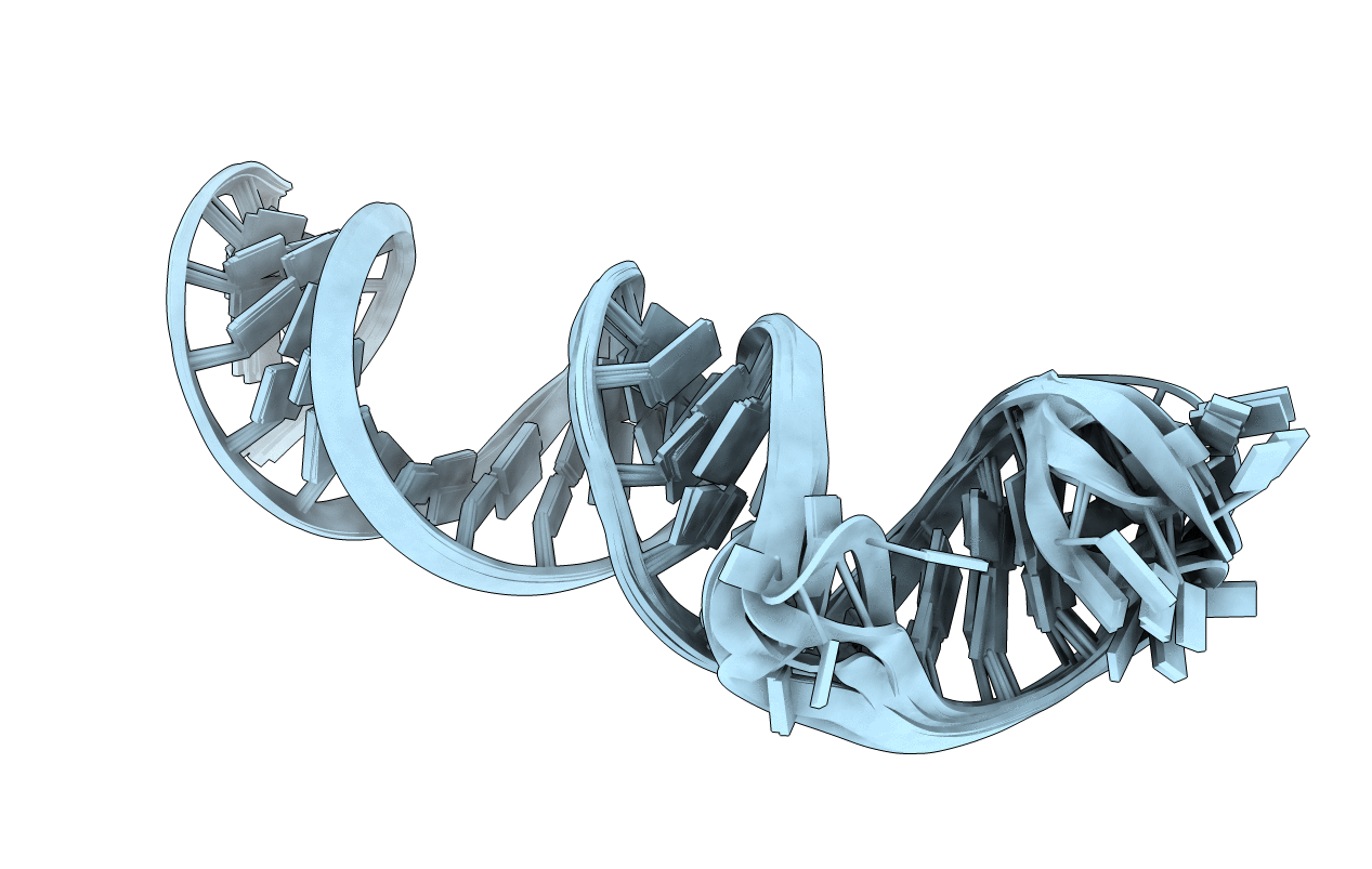
2N4L image
Deposition Date 2015-06-23
Release Date 2015-12-02
Last Version Date 2024-05-01
Entry Detail
PDB ID:
2N4L
Keywords:
Title:
Solution Structure of the HIV-1 Intron Splicing Silencer and its Interactions with the UP1 Domain of hnRNP A1
Biological Source:
Source Organism:
Method Details:
Experimental Method:
Conformers Calculated:
256
Conformers Submitted:
10
Selection Criteria:
structures with the least restraint violations
Macromolecular Entities
Polymer Type:polyribonucleotide
Molecule:RNA (53-MER)
Chain IDs:A
Chain Length:53
Number of Molecules:1
Biological Source:
Ligand Molecules
Primary Citation
Solution Structure of the HIV-1 Intron Splicing Silencer and Its Interactions with the UP1 Domain of Heterogeneous Nuclear Ribonucleoprotein (hnRNP) A1.
J.Biol.Chem. 291 2331 2344 (2016)
PMID: 26607354 DOI: 10.1074/jbc.M115.674564

Abstact

Splicing patterns in human immunodeficiency virus type 1 (HIV-1) are maintained through cis regulatory elements that recruit antagonistic host RNA-binding proteins. The activity of the 3' acceptor site A7 is tightly regulated through a complex network of an intronic splicing silencer (ISS), a bipartite exonic splicing silencer (ESS3a/b), and an exonic splicing enhancer (ESE3). Because HIV-1 splicing depends on protein-RNA interactions, it is important to know the tertiary structures surrounding the splice sites. Herein, we present the NMR solution structure of the phylogenetically conserved ISS stem loop. ISS adopts a stable structure consisting of conserved UG wobble pairs, a folded 2X2 (GU/UA) internal loop, a UU bulge, and a flexible AGUGA apical loop. Calorimetric and biochemical titrations indicate that the UP1 domain of heterogeneous nuclear ribonucleoprotein A1 binds the ISS apical loop site-specifically and with nanomolar affinity. Collectively, this work provides additional insights into how HIV-1 uses a conserved RNA structure to commandeer a host RNA-binding protein.

Legend

Protein

Chemical

Disease

Primary Citation of related structures