2N2J image
Deposition Date 2015-05-09
Release Date 2015-06-10
Last Version Date 2024-05-15
Entry Detail
PDB ID:
2N2J
Keywords:
Title:
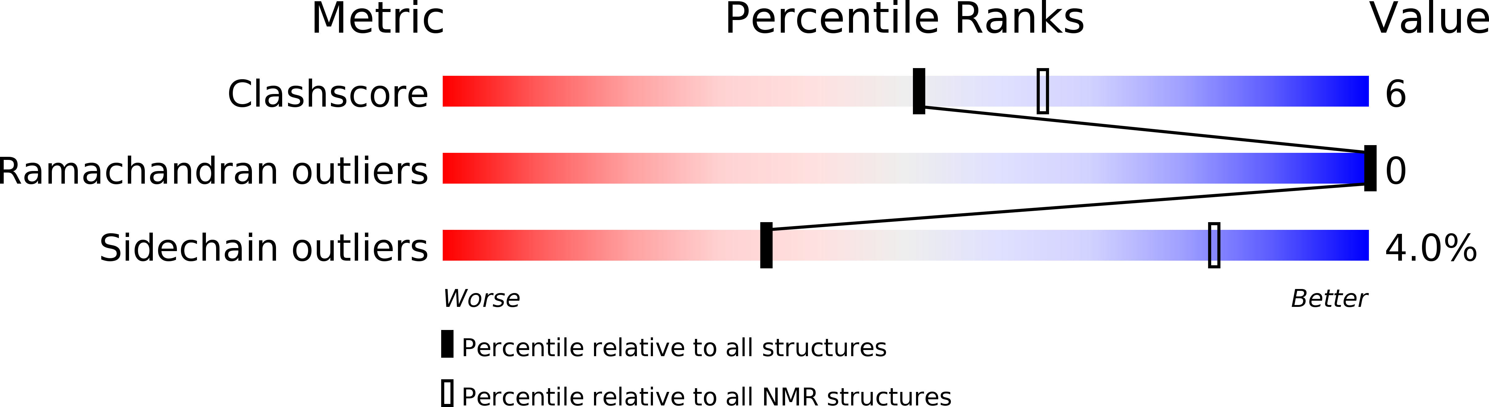
Solution structure of the EBNA-2 N-terminal Dimerization (END) domain from the Epstein-barr virus
Biological Source:
Source Organism:
Host Organism:
Method Details:
Experimental Method:
Conformers Calculated:
100
Conformers Submitted:
10
Selection Criteria:
structures with the lowest energy
Macromolecular Entities
Polymer Type:polypeptide(L)
Molecule:Epstein-Barr nuclear antigen 2
Gene (Uniprot):EBNA2
Chain IDs:A, B
Chain Length:62
Number of Molecules:2
Biological Source:Human herpesvirus 4
Ligand Molecules
Primary Citation
The EBNA-2 N-Terminal Transactivation Domain Folds into a Dimeric Structure Required for Target Gene Activation.
Plos Pathog. 11 e1004910 e1004910 (2015)
PMID: 26024477 DOI: 10.1371/journal.ppat.1004910

Abstact

Epstein-Barr virus (EBV) is a γ-herpesvirus that may cause infectious mononucleosis in young adults. In addition, epidemiological and molecular evidence links EBV to the pathogenesis of lymphoid and epithelial malignancies. EBV has the unique ability to transform resting B cells into permanently proliferating, latently infected lymphoblastoid cell lines. Epstein-Barr virus nuclear antigen 2 (EBNA-2) is a key regulator of viral and cellular gene expression for this transformation process. The N-terminal region of EBNA-2 comprising residues 1-58 appears to mediate multiple molecular functions including self-association and transactivation. However, it remains to be determined if the N-terminus of EBNA-2 directly provides these functions or if these activities merely depend on the dimerization involving the N-terminal domain. To address this issue, we determined the three-dimensional structure of the EBNA-2 N-terminal dimerization (END) domain by heteronuclear NMR-spectroscopy. The END domain monomer comprises a small fold of four β-strands and an α-helix which form a parallel dimer by interaction of two β-strands from each protomer. A structure-guided mutational analysis showed that hydrophobic residues in the dimer interface are required for self-association in vitro. Importantly, these interface mutants also displayed severely impaired self-association and transactivation in vivo. Moreover, mutations of solvent-exposed residues or deletion of the α-helix do not impair dimerization but strongly affect the functional activity, suggesting that the EBNA-2 dimer presents a surface that mediates functionally important intra- and/or intermolecular interactions. Our study shows that the END domain is a novel dimerization fold that is essential for functional activity. Since this specific fold is a unique feature of EBNA-2 it might provide a novel target for anti-viral therapeutics.

Legend

Protein

Chemical

Disease

Primary Citation of related structures