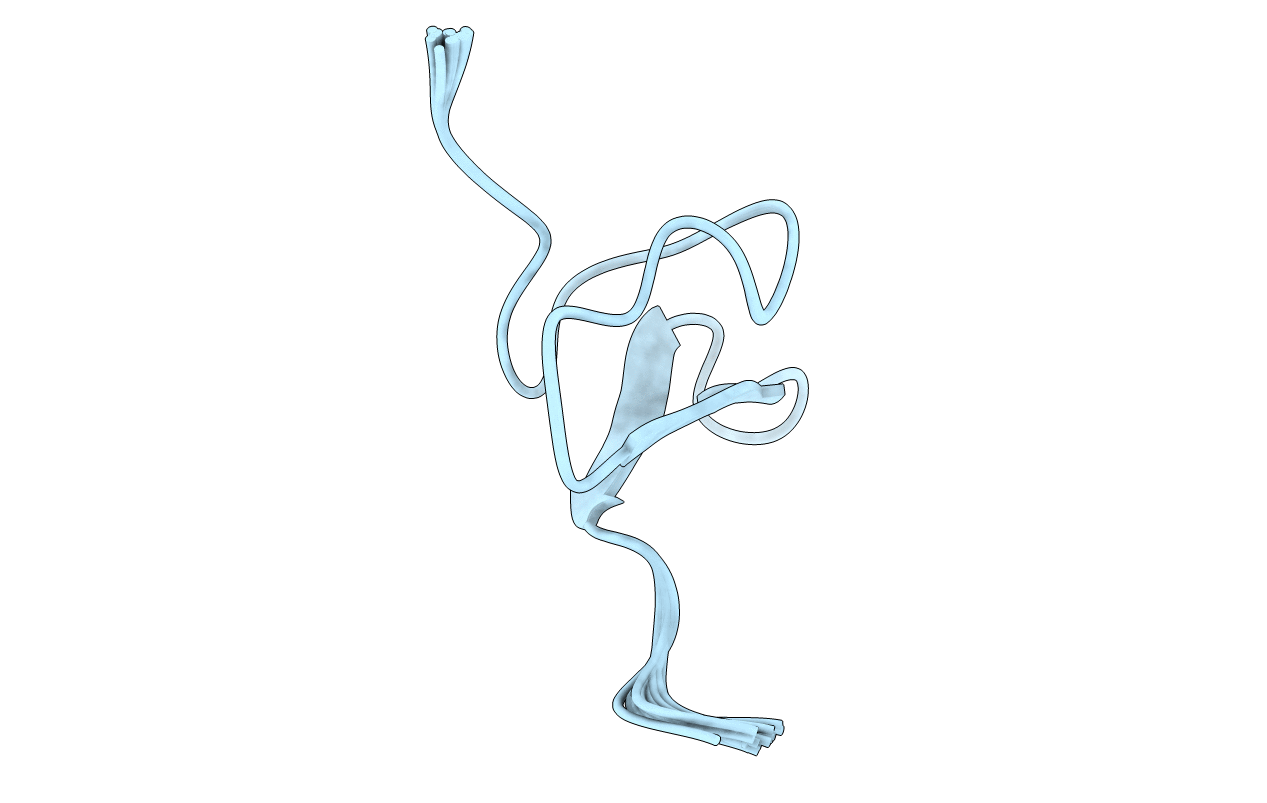
2N1N image
Deposition Date 2015-04-12
Release Date 2016-03-02
Last Version Date 2024-10-09
Entry Detail
PDB ID:
2N1N
Keywords:
Title:
Solution structure of VSTx1
Biological Source:
Source Organism:
Host Organism:
Method Details:
Experimental Method:
Conformers Calculated:
200
Conformers Submitted:
20
Selection Criteria:
target function
Macromolecular Entities
Structures with similar UniProt ID
Protein Blast
Polymer Type:polypeptide(L)
Molecule:Kappa-theraphotoxin-Gr3a
Chain IDs:A
Chain Length:35
Number of Molecules:1
Biological Source:Grammostola rosea
Ligand Molecules
Primary Citation
Molecular basis of the interaction between gating modifier spider toxins and the voltage sensor of voltage-gated ion channels.
Sci Rep 6 34333 34333 (?)
PMID: 27677715 DOI: 10.1038/srep34333

Abstact

Voltage-sensor domains (VSDs) are modular transmembrane domains of voltage-gated ion channels that respond to changes in membrane potential by undergoing conformational changes that are coupled to gating of the ion-conducting pore. Most spider-venom peptides function as gating modifiers by binding to the VSDs of voltage-gated channels and trapping them in a closed or open state. To understand the molecular basis underlying this mode of action, we used nuclear magnetic resonance to delineate the atomic details of the interaction between the VSD of the voltage-gated potassium channel KvAP and the spider-venom peptide VSTx1. Our data reveal that the toxin interacts with residues in an aqueous cleft formed between the extracellular S1-S2 and S3-S4 loops of the VSD whilst maintaining lipid interactions in the gaps formed between the S1-S4 and S2-S3 helices. The resulting network of interactions increases the energetic barrier to the conformational changes required for channel gating, and we propose that this is the mechanism by which gating modifier toxins inhibit voltage-gated ion channels.

Legend

Protein

Chemical

Disease

Primary Citation of related structures