
Deposition Date
2015-04-04
Release Date
2015-11-11
Last Version Date
2024-05-01
Entry Detail
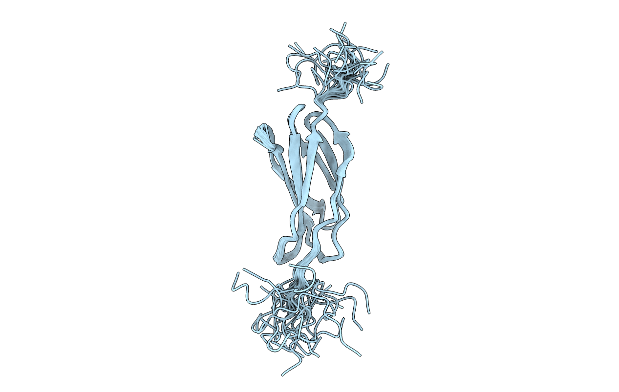
PDB ID:
2N1K
Keywords:
Title:
Structure of the Third Type III Domain from Human Fibronectin
Biological Source:
Source Organism(s):
Homo sapiens (Taxon ID: 9606)
Expression System(s):
Method Details:
Experimental Method:
Conformers Calculated:
80
Conformers Submitted:
25
Selection Criteria:
structures with the lowest energy
