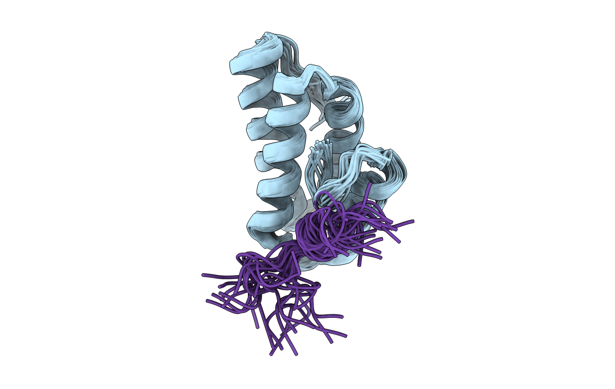
2N1G image
Deposition Date 2015-04-01
Release Date 2016-04-13
Last Version Date 2024-05-01
Entry Detail
PDB ID:
2N1G
Keywords:
Title:
Structure of C-terminal domain of human polymerase Rev1 in complex with PolD3 RIR-motif
Biological Source:
Source Organism:
Homo sapiens (Taxon ID: 9606)
Host Organism:
Method Details:
Experimental Method:
Conformers Calculated:
200
Conformers Submitted:
20
Selection Criteria:
structures with the least restraint violations
Macromolecular Entities
Structures with similar UniProt ID
Protein Blast
Polymer Type:polypeptide(L)
Molecule:DNA repair protein REV1
Gene (Uniprot):REV1
Chain IDs:A
Chain Length:94
Number of Molecules:1
Biological Source:Homo sapiens
Structures with similar UniProt ID
Protein Blast
Polymer Type:polypeptide(L)
Molecule:DNA polymerase delta subunit 3
Gene (Uniprot):POLD3
Chain IDs:B
Chain Length:16
Number of Molecules:1
Biological Source:Homo sapiens
Ligand Molecules
Primary Citation
Interaction between the Rev1 C-Terminal Domain and the PolD3 Subunit of Pol zeta Suggests a Mechanism of Polymerase Exchange upon Rev1/Pol zeta-Dependent Translesion Synthesis.
Biochemistry 55 2043 2053 (2016)
PMID: 26982350 DOI: 10.1021/acs.biochem.5b01282

Abstact

Translesion synthesis (TLS) is a mutagenic branch of cellular DNA damage tolerance that enables bypass replication over DNA lesions carried out by specialized low-fidelity DNA polymerases. The replicative bypass of most types of DNA damage is performed in a two-step process of Rev1/Polζ-dependent TLS. In the first step, a Y-family TLS enzyme, typically Polη, Polι, or Polκ, inserts a nucleotide across a DNA lesion. In the second step, a four-subunit B-family DNA polymerase Polζ (Rev3/Rev7/PolD2/PolD3 complex) extends the distorted DNA primer-template. The coordinated action of error-prone TLS enzymes is regulated through their interactions with the two scaffold proteins, the sliding clamp PCNA and the TLS polymerase Rev1. Rev1 interactions with all other TLS enzymes are mediated by its C-terminal domain (Rev1-CT), which can simultaneously bind the Rev7 subunit of Polζ and Rev1-interacting regions (RIRs) from Polη, Polι, or Polκ. In this work, we identified a previously unknown RIR motif in the C-terminal part of PolD3 subunit of Polζ whose interaction with the Rev1-CT is among the tightest mediated by RIR motifs. Three-dimensional structure of the Rev1-CT/PolD3-RIR complex determined by NMR spectroscopy revealed a structural basis for the relatively high affinity of this interaction. The unexpected discovery of PolD3-RIR motif suggests a mechanism of "inserter" to "extender" DNA polymerase switch upon Rev1/Polζ-dependent TLS, in which the PolD3-RIR binding to the Rev1-CT (i) helps displace the "inserter" Polη, Polι, or Polκ from its complex with Rev1, and (ii) facilitates assembly of the four-subunit "extender" Polζ through simultaneous interaction of Rev1-CT with Rev7 and PolD3 subunits.

Legend

Protein

Chemical

Disease

Primary Citation of related structures