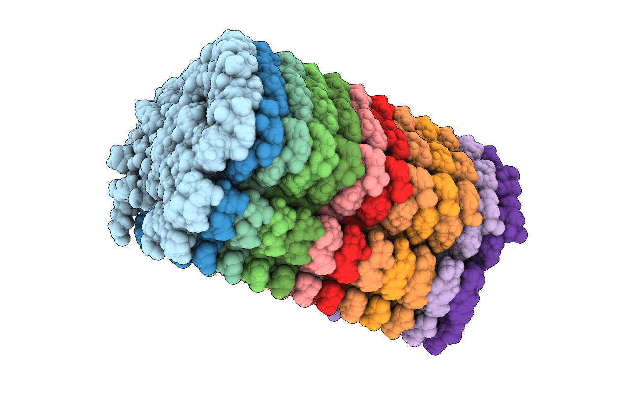
2MXU image
Entry Detail
PDB ID:
2MXU
Keywords:
Title:
42-Residue Beta Amyloid Fibril
Biological Source:
Source Organism:
PDB Version:
Deposition Date:
2015-01-14
Release Date:
2015-05-06
Method Details:
Experimental Method:
Conformers Calculated:
1000
Conformers Submitted:
10
Selection Criteria:
structures with the least restraint violations
Macromolecular Entities
Polymer Type:polypeptide(L)
Description:Amyloid beta A4 protein
Chain IDs:A, B, C, D, E, F, G, H, I, J, K, L
Chain Length:42
Number of Molecules:12
Biological Source:Homo sapiens
Ligand Molecules
Primary Citation
A beta (1-42) fibril structure illuminates self-recognition and replication of amyloid in Alzheimer's disease.
Nat.Struct.Mol.Biol. 22 499 505 (2015)
PMID: 25938662 DOI: 10.1038/nsmb.2991

Abstact

Increasing evidence has suggested that formation and propagation of misfolded aggregates of 42-residue human amyloid β (Aβ(1-42)), rather than of the more abundant Aβ(1-40), provokes the Alzheimer's disease cascade. However, structural details of misfolded Aβ(1-42) have remained elusive. Here we present the atomic model of an Aβ(1-42) amyloid fibril, from solid-state NMR (ssNMR) data. It displays triple parallel-β-sheet segments that differ from reported structures of Aβ(1-40) fibrils. Remarkably, Aβ(1-40) is incompatible with the triple-β-motif, because seeding with Aβ(1-42) fibrils does not promote conversion of monomeric Aβ(1-40) into fibrils via cross-replication. ssNMR experiments suggest that C-terminal Ala42, absent in Aβ(1-40), forms a salt bridge with Lys28 to create a self-recognition molecular switch that excludes Aβ(1-40). The results provide insight into the Aβ(1-42)-selective self-replicating amyloid-propagation machinery in early-stage Alzheimer's disease.

Legend

Protein

Chemical

Disease

Primary Citation of related structures