2MLF image
Deposition Date 2014-02-26
Release Date 2014-03-12
Last Version Date 2024-05-15
Entry Detail
PDB ID:
2MLF
Title:
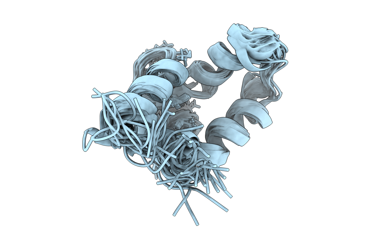
NMR structure of the dilated cardiomyopathy mutation G159D in troponin C bound to the anchoring region of troponin I
Biological Source:
Source Organism:
Homo sapiens (Taxon ID: 9606)
Host Organism:
Method Details:
Experimental Method:
Conformers Calculated:
50
Conformers Submitted:
20
Selection Criteria:
target function
Macromolecular Entities
Polymer Type:polypeptide(L)
Molecule:Troponin C, slow skeletal and cardiac muscles
Gene (Uniprot):TNNC1
Chain IDs:A (auth: C)
Chain Length:72
Number of Molecules:1
Biological Source:Homo sapiens
Ligand Molecules
Primary Citation
The dilated cardiomyopathy G159D mutation in cardiac troponin C weakens the anchoring interaction with troponin I.
Biochemistry 47 10950 10960 (2008)
PMID: 18803402 DOI: 10.1021/bi801165c

Abstact

NMR spectroscopy has been employed to elucidate the molecular consequences of the DCM G159D mutation on the structure and dynamics of troponin C, and its interaction with troponin I (TnI). Since the molecular effects of human mutations are often subtle, all NMR experiments were conducted as direct side-by-side comparisons of the wild-type C-domain of troponin C (cCTnC) and the mutant protein, G159D. With the mutation, the affinity toward the anchoring region of cTnI (cTnI 34-71) was reduced (K D = 3.0 +/- 0.6 microM) compared to that of the wild type (K D < 1 microM). Overall, the structure and dynamics of the G159D.cTnI 34-71 complex were very similar to those of the cCTnC.cTnI 34-71 complex. There were, however, significant changes in the (1)H, (13)C, and (15)N NMR chemical shifts, especially for the residues in direct contact with cTnI 34-71, and the changes in NOE connectivity patterns between the G159D.cTnI 34-71 and cCTnC.cTnI 34-71 complexes. Thus, the most parsimonious hypothesis is that the development of disease results from the poor anchoring of cTnI to cCTnC, with the resulting increase in the level of acto-myosin inhibition in agreement with physiological data. Another possibility is that long-range electrostatic interactions affect the binding of the inhibitory and switch regions of cTnI (cTnI 128-147 and cTnI 147-163) and/or the cardiac specific N-terminus of cTnI (cTnI 1-29) to the N-domain of cTnC. These important interactions are all spatially close in the X-ray structure of the cardiac TnC core.

Legend

Protein

Chemical

Disease

Primary Citation of related structures