Deposition Date
2014-02-13
Release Date
2014-06-18
Last Version Date
2024-11-06
Entry Detail
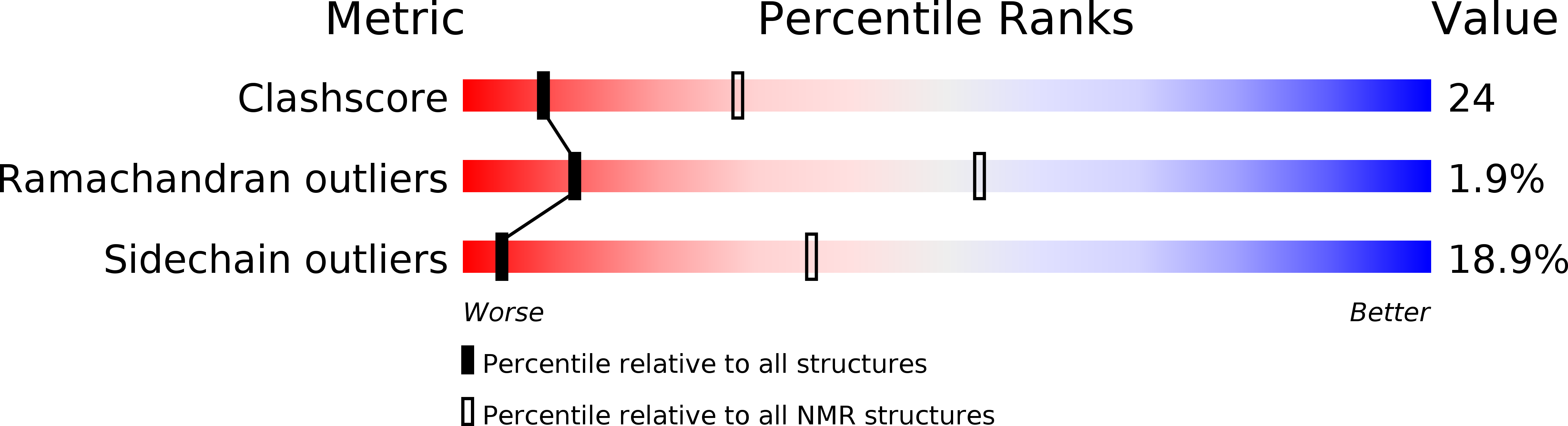
PDB ID:
2MKW
Keywords:
Title:
Solution Structure of 6aJl2 and 6aJL2-R24G Amyloidogenics Light Chain Proteins
Biological Source:
Source Organism(s):
Homo sapiens (Taxon ID: 9606)
Expression System(s):
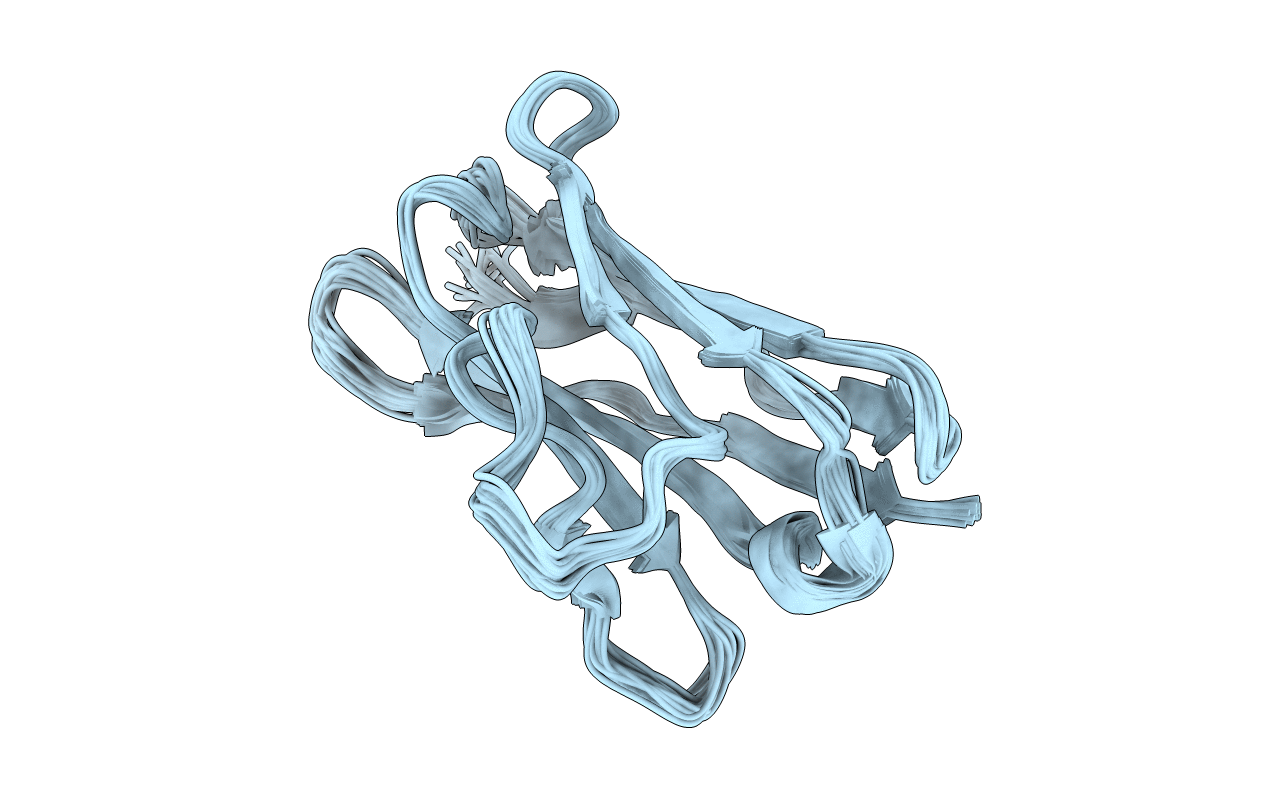
Method Details:
Experimental Method:
Conformers Calculated:
100
Conformers Submitted:
20
Selection Criteria:
structures with the lowest energy