2M8D image
Deposition Date 2013-05-17
Release Date 2013-07-10
Last Version Date 2024-05-15
Entry Detail
PDB ID:
2M8D
Title:
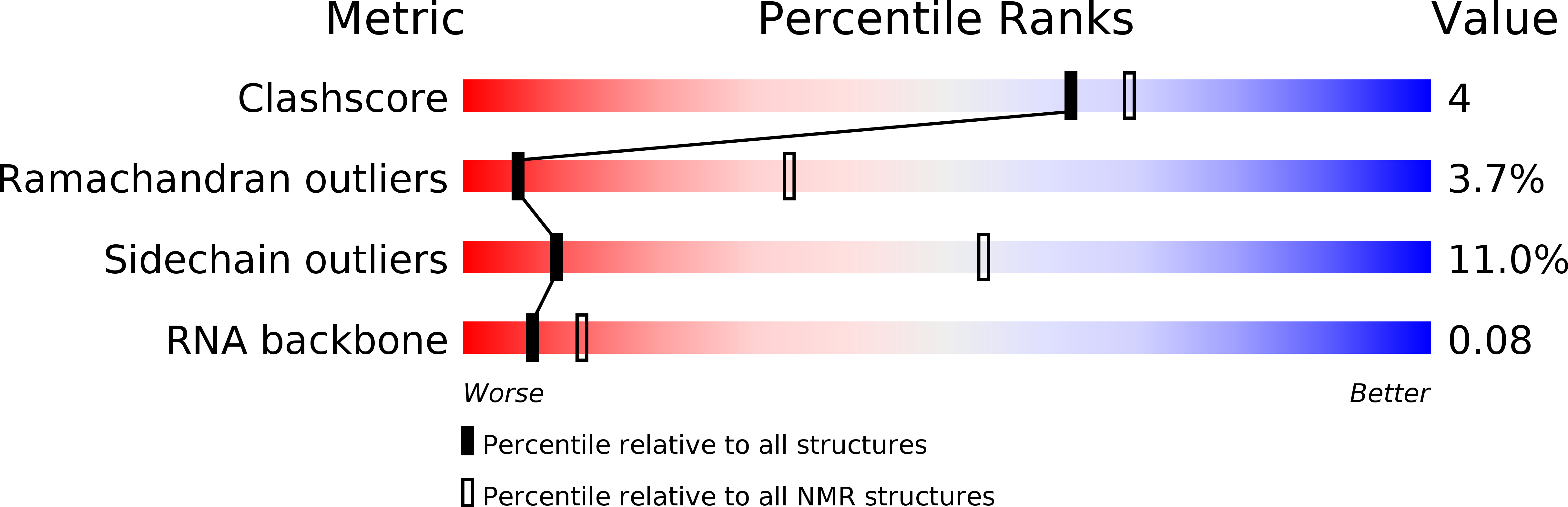
Structure of SRSF1 RRM2 in complex with the RNA 5'-UGAAGGAC-3'
Biological Source:
Source Organism(s):
Homo sapiens (Taxon ID: 9606)
Expression System(s):
Method Details:
Experimental Method:
Conformers Calculated:
50
Conformers Submitted:
16
Selection Criteria:
structures with the lowest energy
Macromolecular Entities
Structures with similar UniProt ID
Protein Blast
Polymer Type:polypeptide(L)
Molecule:Serine/arginine-rich splicing factor 1
Gene (Uniprot):SRSF1
Chain IDs:B
Chain Length:91
Number of Molecules:1
Biological Source:Homo sapiens
Ligand Molecules
Primary Citation
Isolated pseudo-RNA-recognition motifs of SR proteins can regulate splicing using a noncanonical mode of RNA recognition.
Proc.Natl.Acad.Sci.USA 110 E2802 E2811 (2013)
PMID: 23836656 DOI: 10.1073/pnas.1303445110

Abstact

Serine/arginine (SR) proteins, one of the major families of alternative-splicing regulators in Eukarya, have two types of RNA-recognition motifs (RRMs): a canonical RRM and a pseudo-RRM. Although pseudo-RRMs are crucial for activity of SR proteins, their mode of action was unknown. By solving the structure of the human SRSF1 pseudo-RRM bound to RNA, we discovered a very unusual and sequence-specific RNA-binding mode that is centered on one α-helix and does not involve the β-sheet surface, which typically mediates RNA binding by RRMs. Remarkably, this mode of binding is conserved in all pseudo-RRMs tested. Furthermore, the isolated pseudo-RRM is sufficient to regulate splicing of about half of the SRSF1 target genes tested, and the bound α-helix is a pivotal element for this function. Our results strongly suggest that SR proteins with a pseudo-RRM frequently regulate splicing by competing with, rather than recruiting, spliceosome components, using solely this unusual RRM.

Legend

Protein

Chemical

Disease

Primary Citation of related structures
Feedback Form
Name
Email
Institute
Feedback