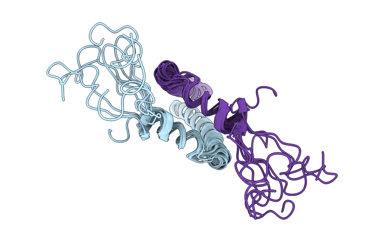
2M20 image
Entry Detail
PDB ID:
2M20
Title:
EGFR transmembrane - juxtamembrane (TM-JM) segment in bicelles: MD guided NMR refined structure.
Biological Source:
Source Organism:
Host Organism:
PDB Version:
Deposition Date:
2012-12-11
Release Date:
2013-02-20
Method Details:
Experimental Method:
Conformers Calculated:
50
Conformers Submitted:
10
Selection Criteria:
structures with the lowest energy
Macromolecular Entities
Polymer Type:polypeptide(L)
Description:Epidermal growth factor receptor
Mutations:M9L (M650 in UNP P00533), M27I (M668 in UNP P00533)
Chain IDs:A, B
Chain Length:60
Number of Molecules:2
Biological Source:Homo sapiens
Ligand Molecules
Primary Citation
Conformational Coupling across the Plasma Membrane in Activation of the EGF Receptor.
Cell(Cambridge,Mass.) 152 543 556 (2013)
PMID: 23374349 DOI: 10.1016/j.cell.2012.12.032

Abstact

How the epidermal growth factor receptor (EGFR) activates is incompletely understood. The intracellular portion of the receptor is intrinsically active in solution, and to study its regulation, we measured autophosphorylation as a function of EGFR surface density in cells. Without EGF, intact EGFR escapes inhibition only at high surface densities. Although the transmembrane helix and the intracellular module together suffice for constitutive activity even at low densities, the intracellular module is inactivated when tethered on its own to the plasma membrane, and fluorescence cross-correlation shows that it fails to dimerize. NMR and functional data indicate that activation requires an N-terminal interaction between the transmembrane helices, which promotes an antiparallel interaction between juxtamembrane segments and release of inhibition by the membrane. We conclude that EGF binding removes steric constraints in the extracellular module, promoting activation through N-terminal association of the transmembrane helices.

Legend

Protein

Chemical

Disease

Primary Citation of related structures