2LX4 image
Deposition Date 2012-08-14
Release Date 2013-01-09
Last Version Date 2024-05-01
Entry Detail
PDB ID:
2LX4
Title:
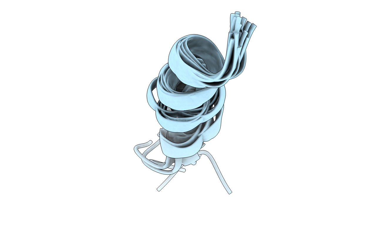
NMR solution structure of peptide a2N(1-17) from Mus musculus V-ATPase
Biological Source:
Source Organism:
Mus musculus (Taxon ID: 10090)
Method Details:
Experimental Method:
Conformers Calculated:
20
Conformers Submitted:
10
Selection Criteria:
structures with the lowest energy
Macromolecular Entities
Structures with similar UniProt ID
Protein Blast
Polymer Type:polypeptide(L)
Molecule:V-type proton ATPase 116 kDa subunit a isoform 2
Gene (Uniprot):Atp6v0a2
Chain IDs:A
Chain Length:17
Number of Molecules:1
Biological Source:Mus musculus
Ligand Molecules
Primary Citation
The N Termini of a-Subunit Isoforms Are Involved in Signaling between Vacuolar H+-ATPase (V-ATPase) and Cytohesin-2.
J.Biol.Chem. 288 5896 5913 (2013)
PMID: 23288846 DOI: 10.1074/jbc.M112.409169

Abstact

Previously, we reported an acidification-dependent interaction of the endosomal vacuolar H(+)-ATPase (V-ATPase) with cytohesin-2, a GDP/GTP exchange factor (GEF), suggesting that it functions as a pH-sensing receptor. Here, we have studied the molecular mechanism of signaling between the V-ATPase, cytohesin-2, and Arf GTP-binding proteins. We found that part of the N-terminal cytosolic tail of the V-ATPase a2-subunit (a2N), corresponding to its first 17 amino acids (a2N(1-17)), potently modulates the enzymatic GDP/GTP exchange activity of cytohesin-2. Moreover, this peptide strongly inhibits GEF activity via direct interaction with the Sec7 domain of cytohesin-2. The structure of a2N(1-17) and its amino acids Phe(5), Met(10), and Gln(14) involved in interaction with Sec7 domain were determined by NMR spectroscopy analysis. In silico docking experiments revealed that part of the V-ATPase formed by its a2N(1-17) epitope competes with the switch 2 region of Arf1 and Arf6 for binding to the Sec7 domain of cytohesin-2. The amino acid sequence alignment and GEF activity studies also uncovered the conserved character of signaling between all four (a1-a4) a-subunit isoforms of mammalian V-ATPase and cytohesin-2. Moreover, the conserved character of this phenomenon was also confirmed in experiments showing binding of mammalian cytohesin-2 to the intact yeast V-ATPase holo-complex. Thus, here we have uncovered an evolutionarily conserved function of the V-ATPase as a novel cytohesin-signaling receptor.

Legend

Protein

Chemical

Disease

Primary Citation of related structures