2LWR image
Entry Detail
PDB ID:
2LWR
Keywords:
Title:
Solution Structure of RING2 Domain from Parkin
Biological Source:
Source Organism:
Host Organism:
PDB Version:
Deposition Date:
2012-08-06
Release Date:
2013-06-12
Method Details:
Experimental Method:
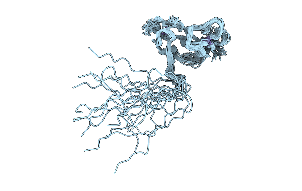
Conformers Calculated:
20
Conformers Submitted:
19
Selection Criteria:
structures with the least restraint violations
Macromolecular Entities
Polymer Type:polypeptide(L)
Description:SD01679p
Chain IDs:A
Chain Length:68
Number of Molecules:1
Biological Source:Drosophila melanogaster
Ligand Molecules
Primary Citation
A molecular explanation for the recessive nature of parkin-linked Parkinson's disease.
Nat Commun 4 1983 1983 (2013)
PMID: 23770917 DOI: 10.1038/ncomms2983

Abstact

Mutations in the park2 gene, encoding the RING-inBetweenRING-RING E3 ubiquitin ligase parkin, cause 50% of autosomal recessive juvenile Parkinsonism cases. More than 70 known pathogenic mutations occur throughout parkin, many of which cluster in the inhibitory amino-terminal ubiquitin-like domain, and the carboxy-terminal RING2 domain that is indispensable for ubiquitin transfer. A structural rationale showing how autosomal recessive juvenile Parkinsonism mutations alter parkin function is still lacking. Here we show that the structure of parkin RING2 is distinct from canonical RING E3 ligases and lacks key elements required for E2-conjugating enzyme recruitment. Several pathogenic mutations in RING2 alter the environment of a single surface-exposed catalytic cysteine to inhibit ubiquitination. Native parkin adopts a globular inhibited conformation in solution facilitated by the association of the ubiquitin-like domain with the RING-inBetweenRING-RING C-terminus. Autosomal recessive juvenile Parkinsonism mutations disrupt this conformation. Finally, parkin autoubiquitinates only in cis, providing a molecular explanation for the recessive nature of autosomal recessive juvenile Parkinsonism.

Legend

Protein

Chemical

Disease

Primary Citation of related structures