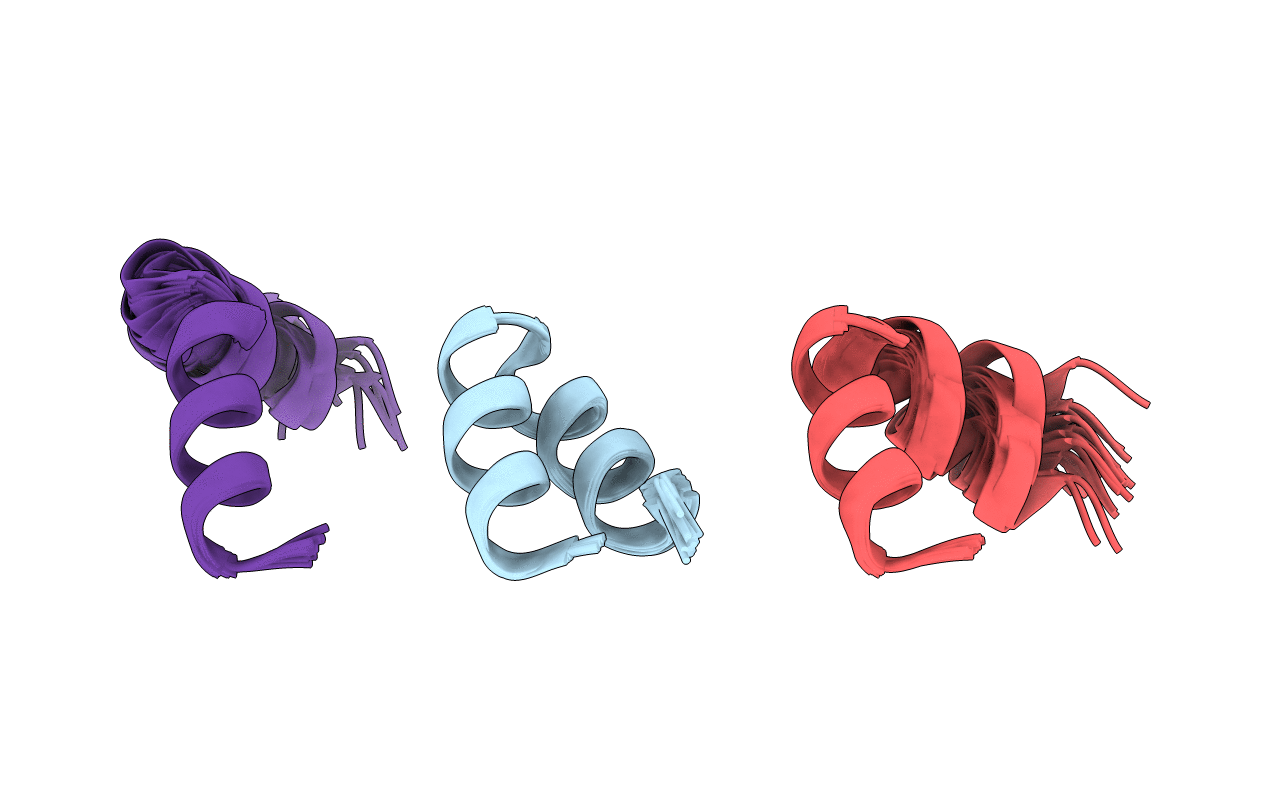
2LWA image
Deposition Date 2012-07-26
Release Date 2012-12-05
Last Version Date 2024-05-15
Entry Detail
PDB ID:
2LWA
Title:
Conformational ensemble for the G8A mutant of the influenza hemagglutinin fusion peptide
Biological Source:
Source Organism:
Host Organism:
Method Details:
Experimental Method:
Conformers Calculated:
480
Conformers Submitted:
20
Selection Criteria:
structures with the lowest energy
Macromolecular Entities
Structures with similar UniProt ID
Protein Blast
Polymer Type:polypeptide(L)
Molecule:HEMAGGLUTININ FUSION PEPTIDE G8A MUTANT
Gene (Uniprot):HA
Mutagens:G8A
Chain IDs:A, B, C
Chain Length:30
Number of Molecules:3
Biological Source:Influenza A virus
Ligand Molecules
Primary Citation
pH-triggered, activated-state conformations of the influenza hemagglutinin fusion peptide revealed by NMR.
Proc.Natl.Acad.Sci.USA 109 19994 19999 (2012)
PMID: 23169643 DOI: 10.1073/pnas.1213801109

Abstact

The highly conserved first 23 residues of the influenza hemagglutinin HA2 subunit constitute the fusion domain, which plays a pivotal role in fusing viral and host-cell membranes. At neutral pH, this peptide adopts a tight helical hairpin wedge structure, stabilized by aliphatic hydrogen bonding and charge-dipole interactions. We demonstrate that at low pH, where the fusion process is triggered, the native peptide transiently visits activated states that are very similar to those sampled by a G8A mutant. This mutant retains a small fraction of helical hairpin conformation, in rapid equilibrium with at least two open structures. The exchange rate between the closed and open conformations of the wild-type fusion peptide is ~40 kHz, with a total open-state population of ~20%. Transitions to these activated states are likely to play a crucial role in formation of the fusion pore, an essential structure required in the final stage of membrane fusion.

Legend

Protein

Chemical

Disease

Primary Citation of related structures