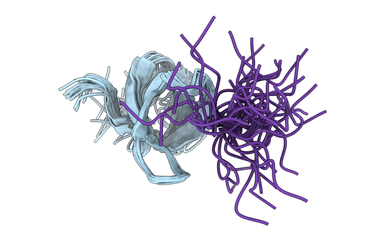
2LTO image
Entry Detail
PDB ID:
2LTO
Title:
TDRD3 complex
Biological Source:
Source Organism:
Host Organism:
PDB Version:
Deposition Date:
2012-05-30
Release Date:
2013-01-16
Method Details:
Experimental Method:
Conformers Calculated:
20
Conformers Submitted:
20
Selection Criteria:
target function
Macromolecular Entities
Polymer Type:polypeptide(L)
Description:Tudor domain-containing protein 3
Chain IDs:A
Chain Length:58
Number of Molecules:1
Biological Source:Homo sapiens
Polymer Type:polypeptide(L)
Description:DNA-directed RNA polymerase II subunit RPB1
Chain IDs:B
Chain Length:13
Number of Molecules:1
Biological Source:Homo sapiens
Modified Residue
Compound ID Chain ID Parent Comp ID Details 2D Image
DA2 B ARG NG,NG-DIMETHYL-L-ARGININE
Ligand Molecules
Primary Citation
Recognition of asymmetrically dimethylated arginine by TDRD3.
Nucleic Acids Res. 40 11748 11755 (2012)
PMID: 23066109 DOI: 10.1093/nar/gks929

Abstact

Asymmetric dimethylarginine (aDMA) marks are placed on histones and the C-terminal domain (CTD) of RNA Polymerase II (RNAP II) and serve as a signal for recruitment of appropriate transcription and processing factors in coordination with transcription cycle. In contrast to other Tudor domain-containing proteins, Tudor domain-containing protein 3 (TDRD3) associates selectively with the aDMA marks but not with other methylarginine motifs. Here, we report the solution structure of the Tudor domain of TDRD3 bound to the asymmetrically dimethylated CTD. The structure and mutational analysis provide a molecular basis for how TDRD3 recognizes the aDMA mark. The unique aromatic cavity of the TDRD3 Tudor domain with a tyrosine in position 566 creates a selectivity filter for the aDMA residue. Our work contributes to the understanding of substrate selectivity rules of the Tudor aromatic cavity, which is an important structural motif for reading of methylation marks.

Legend

Protein

Chemical

Disease

Primary Citation of related structures