2LRK image
Deposition Date 2012-04-06
Release Date 2012-05-16
Last Version Date 2024-05-01
Entry Detail
PDB ID:
2LRK
Keywords:
Title:
Solution Structures of the IIA(Chitobiose)-HPr complex of the N,N'-Diacetylchitobiose
Biological Source:
Source Organism:
Host Organism:
Method Details:
Experimental Method:
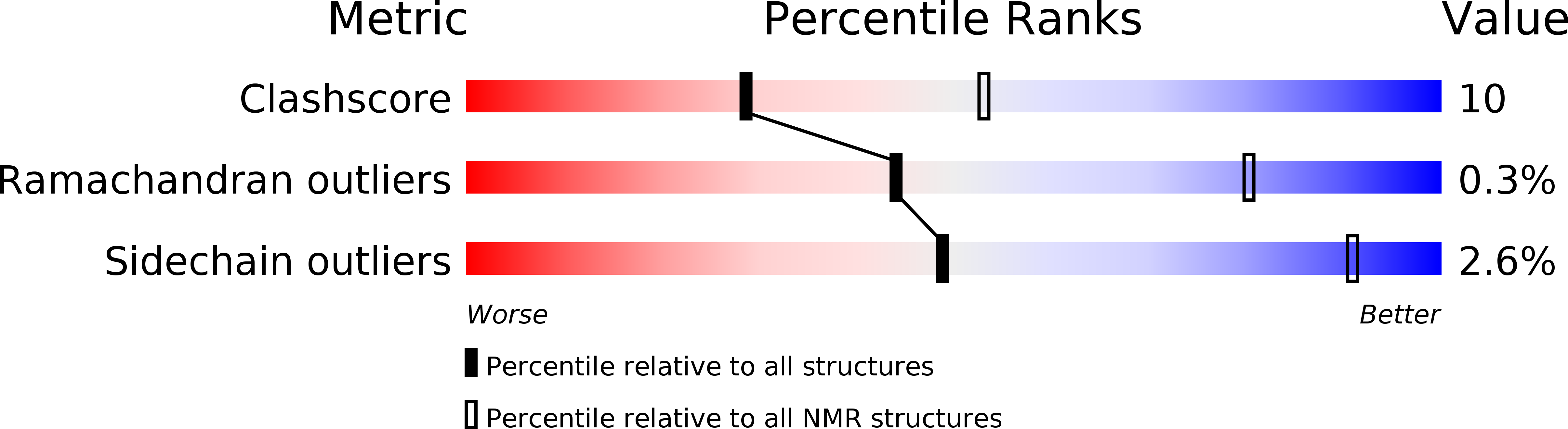
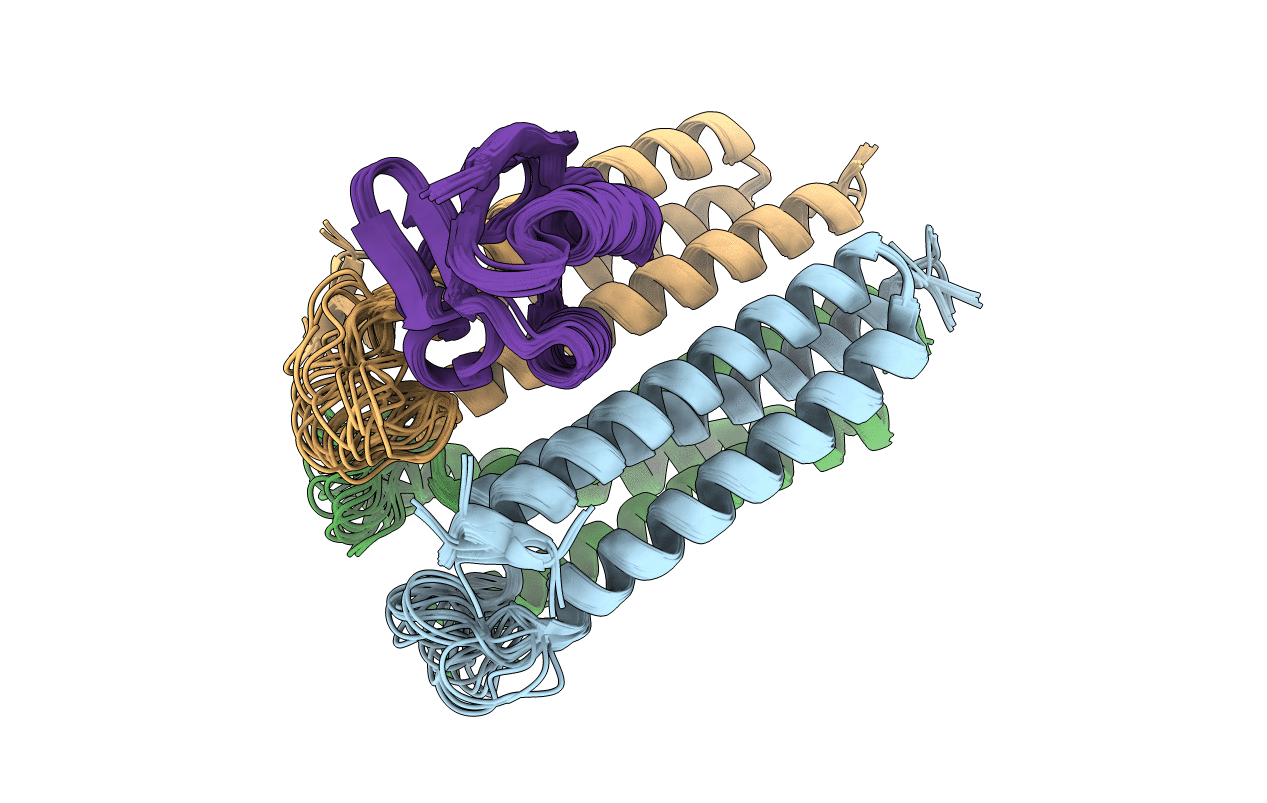
Conformers Calculated:
100
Conformers Submitted:
21
Selection Criteria:
structures with the least restraint violations
Macromolecular Entities
Polymer Type:polypeptide(L)
Molecule:N,N'-diacetylchitobiose-specific phosphotransferase enzyme IIA component
Gene (Uniprot):chbA
Mutations:H76E, D79L
Chain IDs:B (auth: A), C (auth: B), D (auth: C)
Chain Length:103
Number of Molecules:3
Biological Source:Escherichia coli
Polymer Type:polypeptide(L)
Molecule:Phosphocarrier protein HPr
Gene (Uniprot):ptsH
Chain IDs:A (auth: D)
Chain Length:85
Number of Molecules:1
Biological Source:Escherichia coli
Ligand Molecules
Primary Citation
Solution Structure of the IIAChitobiose-HPr Complex of the N,N'-Diacetylchitobiose Branch of the Escherichia coli Phosphotransferase System.
J.Biol.Chem. 287 23819 23829 (2012)
PMID: 22593574 DOI: 10.1074/jbc.M112.371492

Abstact

The solution structure of the complex of enzyme IIA of the N,N'-diacetylchitobiose (Chb) transporter with the histidine phosphocarrier protein HPr has been solved by NMR. The IIA(Chb)-HPr complex completes the structure elucidation of representative cytoplasmic complexes for all four sugar branches of the bacterial phosphoryl transfer system (PTS). The active site His-89 of IIA(Chb) was mutated to Glu to mimic the phosphorylated state. IIA(Chb)(H89E) and HPr form a weak complex with a K(D) of ~0.7 mM. The interacting binding surfaces, concave for IIA(Chb) and convex for HPr, complement each other in terms of shape, residue type, and charge distribution, with predominantly hydrophobic residues, interspersed by some uncharged polar residues, located centrally, and polar and charged residues at the periphery. The active site histidine of HPr, His-15, is buried within the active site cleft of IIA(Chb) formed at the interface of two adjacent subunits of the IIA(Chb) trimer, thereby coming into close proximity with the active site residue, H89E, of IIA(Chb). A His89-P-His-15 pentacoordinate phosphoryl transition state can readily be modeled without necessitating any significant conformational changes, thereby facilitating rapid phosphoryl transfer. Comparison of the IIA(Chb)-HPr complex with the IIA(Chb)-IIB(Chb) complex, as well as with other cytoplasmic complexes of the PTS, highlights a unifying mechanism for recognition of structurally diverse partners. This involves generating similar binding surfaces from entirely different underlying structural elements, large interaction surfaces coupled with extensive redundancy, and side chain conformational plasticity to optimize diverse sets of intermolecular interactions.

Legend

Protein

Chemical

Disease

Primary Citation of related structures