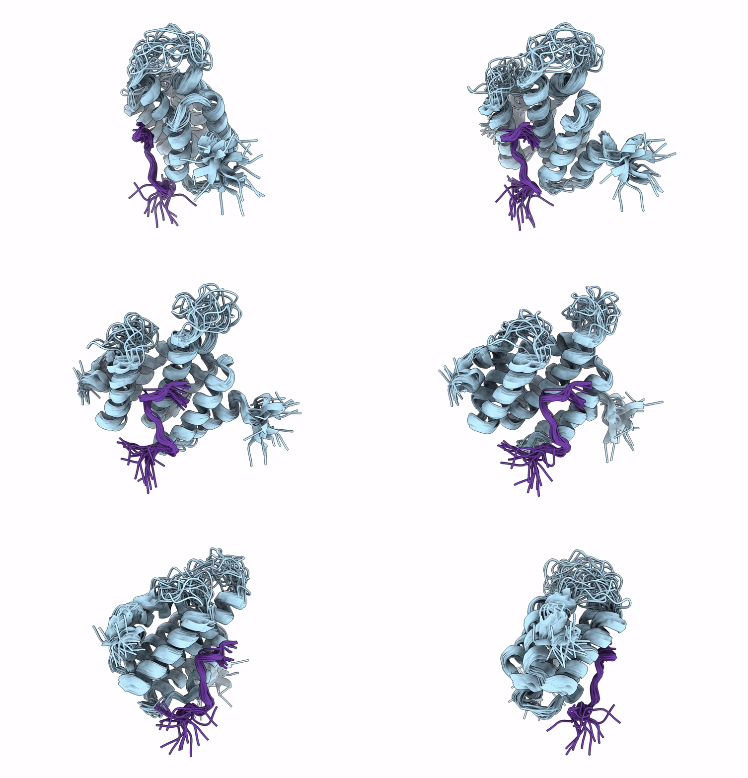
2LO6 image
Deposition Date 2012-01-17
Release Date 2012-12-26
Last Version Date 2024-11-06
Entry Detail
PDB ID:
2LO6
Keywords:
Title:
Structure of Nrd1 CID bound to phosphorylated RNAP II CTD
Biological Source:
Source Organism:
Host Organism:
Method Details:
Experimental Method:
Conformers Calculated:
40
Conformers Submitted:
20
Selection Criteria:
structures with the lowest energy
Macromolecular Entities
Polymer Type:polypeptide(L)
Molecule:Protein NRD1
Gene (Uniprot):NRD1
Chain IDs:A
Chain Length:161
Number of Molecules:1
Biological Source:Saccharomyces cerevisiae S288c
Polymer Type:polypeptide(L)
Molecule:DNA-directed RNA polymerase II subunit RPB1
Gene (Uniprot):RPO21
Chain IDs:B
Chain Length:14
Number of Molecules:1
Biological Source:Saccharomyces cerevisiae S288c
Modified Residue
Compound ID Chain ID Parent Comp ID Details 2D Image
SEP B SER PHOSPHOSERINE
Ligand Molecules
Primary Citation
Serine phosphorylation and proline isomerization in RNAP II CTD control recruitment of Nrd1.
Genes Dev. 26 1891 1896 (2012)
PMID: 22892239 DOI: 10.1101/gad.192781.112

Abstact

Recruitment of appropriate RNA processing factors to the site of transcription is controlled by post-translational modifications of the C-terminal domain (CTD) of RNA polymerase II (RNAP II). Here, we report the solution structure of the Ser5 phosphorylated (pSer5) CTD bound to Nrd1. The structure reveals a direct recognition of pSer5 by Nrd1 that requires the cis conformation of the upstream pSer5-Pro6 peptidyl-prolyl bond of the CTD. Mutations at the complex interface diminish binding affinity and impair processing or degradation of noncoding RNAs. These findings underpin the interplay between covalent and noncovalent changes in the CTD structure that constitute the CTD code.

Legend

Protein

Chemical

Disease

Primary Citation of related structures