2LLX image
Deposition Date 2011-11-18
Release Date 2012-05-30
Last Version Date 2024-05-15
Entry Detail
PDB ID:
2LLX
Keywords:
Title:
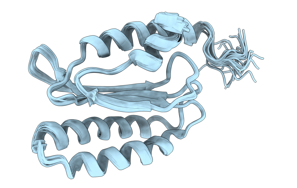
Solution structure of the N-terminal domain of human polypeptide chain release factor eRF1
Biological Source:
Source Organism:
Homo sapiens (Taxon ID: 9606)
Host Organism:
Method Details:
Experimental Method:
Conformers Calculated:
50
Conformers Submitted:
20
Selection Criteria:
structures with the least restraint violations
Macromolecular Entities
Polymer Type:polypeptide(L)
Molecule:Eukaryotic peptide chain release factor subunit 1
Gene (Uniprot):ETF1
Chain IDs:A
Chain Length:150
Number of Molecules:1
Biological Source:Homo sapiens
Ligand Molecules
Primary Citation
Structure and dynamics in solution of the stop codon decoding N-terminal domain of the human polypeptide chain release factor eRF1.
Protein Sci. 21 896 903 (2012)
PMID: 22517631 DOI: 10.1002/pro.2067

Abstact

The high-resolution NMR structure of the N-domain of human eRF1, responsible for stop codon recognition, has been determined in solution. The overall fold of the protein is the same as that found in the crystal structure. However, the structures of several loops, including those participating in stop codon decoding, are different. Analysis of the NMR relaxation data reveals that most of the regions with the highest structural discrepancy between the solution and solid states undergo internal motions on the ps-ns and ms time scales. The NMR data show that the N-domain of human eRF1 exists in two conformational states. The distribution of the residues having the largest chemical shift differences between the two forms indicates that helices α2 and α3, with the NIKS loop between them, can switch their orientation relative to the β-core of the protein. Such structural plasticity may be essential for stop codon recognition by human eRF1.

Legend

Protein

Chemical

Disease

Primary Citation of related structures