2LHM image
Deposition Date 1991-10-02
Release Date 1992-04-15
Last Version Date 2024-11-13
Entry Detail
PDB ID:
2LHM
Title:
CRYSTAL STRUCTURES OF THE APO-AND HOLOMUTANT HUMAN LYSOZYMES WITH AN INTRODUCED CA2+ BINDING SITE
Biological Source:
Source Organism:
Homo sapiens (Taxon ID: 9606)
Method Details:
Experimental Method:
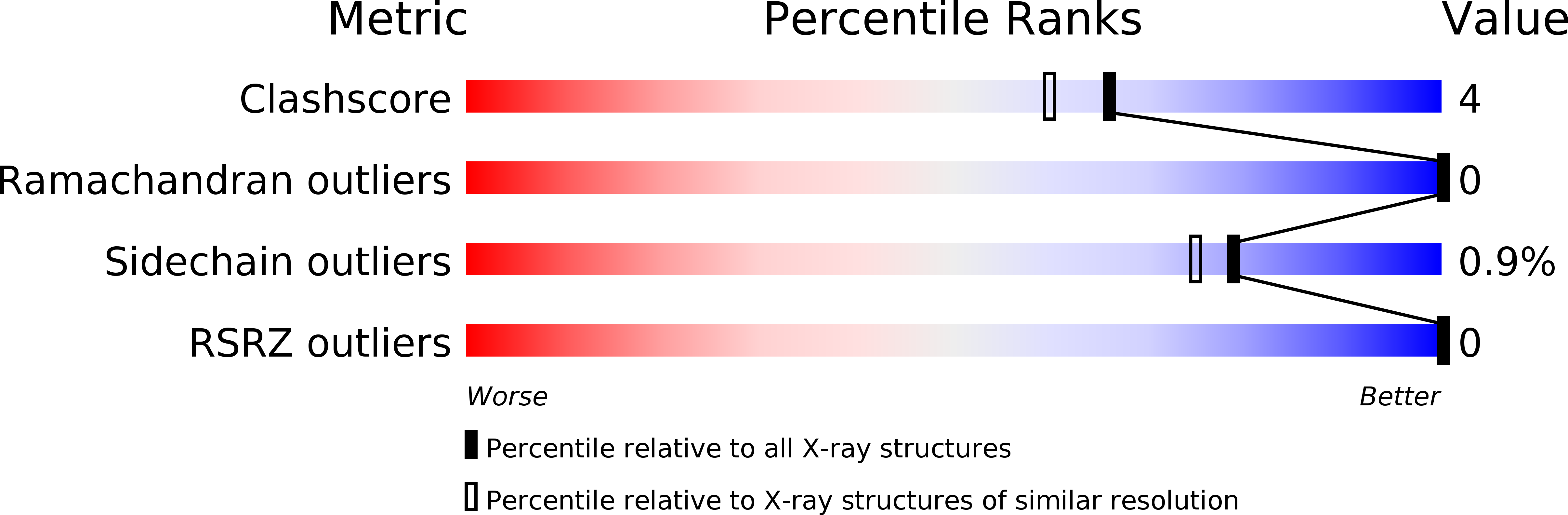
Resolution:
1.80 Å
R-Value Observed:
0.16
Space Group:
P 21 21 21
Macromolecular Entities
Structures with similar UniProt ID
Protein Blast
Polymer Type:polypeptide(L)
Molecule:HUMAN LYSOZYME
Gene (Uniprot):LYZ
Chain IDs:A
Chain Length:130
Number of Molecules:1
Biological Source:Homo sapiens
Primary Citation
Crystal structures of the apo- and holomutant human lysozymes with an introduced Ca2+ binding site.
J.Biol.Chem. 266 20666 20671 (1991)
PMID: 1939116

Abstact

The three-dimensional structures of apo- and holomutant human lysozymes (D86/92 lysozyme), in which a calcium binding site was designed and created for enhancing molecular stability by replacing both Gln86 and Ala92 with aspartic acids, were refined at 1.8-A resolution by x-ray crystallography. The overall structures and crystallographic thermal factors of all three proteins, the apo-, holo-D86/92, and the wild-type human lysozymes, were essentially identical; these results showed that the introduction of the calcium binding site did not affect either the overall structure or molecular rigidity of the proteins. However, structure analyses of the apo-D86/92 lysozyme revealed that the mutations affected the side chain conformation of residue 86 and hydrogen networks between the protein and the internal solvent molecules. In the structure of the holo-D86/92 lysozyme, seven oxygen ligands formed a slightly distorted pentagonal bipyramid around the calcium ion, indicating that the coordination around the calcium ion was quite similar to that in baboon alpha-lactalbumin. The pentagonal bipyramid coordination could be one of the most widely found and appropriate calcium binding schemes in proteins.

Legend

Protein

Chemical

Disease

Primary Citation of related structures
Feedback Form
Name
Email
Institute
Feedback