2LG4 image
Deposition Date 2011-07-20
Release Date 2012-07-25
Last Version Date 2024-11-27
Entry Detail
PDB ID:
2LG4
Title:
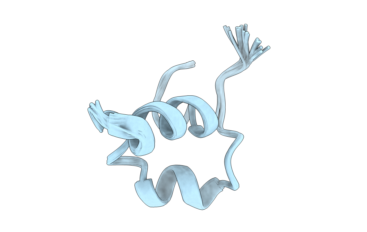
3D solution structure of antimicrobial peptide aurelin
Biological Source:
Source Organism:
Aurelia aurita (Taxon ID: 6145)
Host Organism:
Method Details:
Experimental Method:
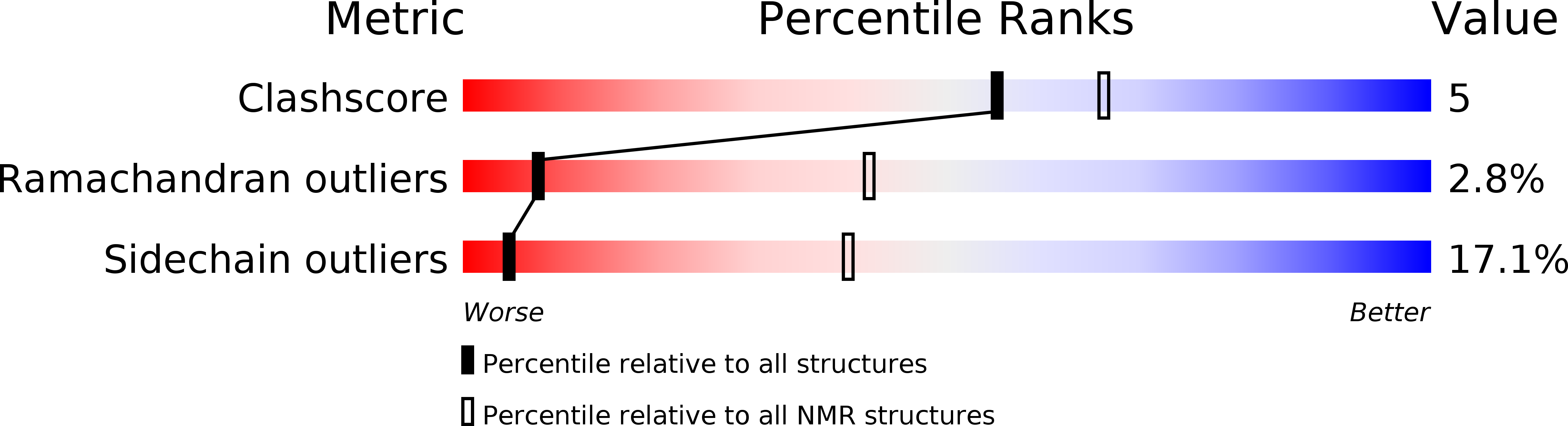
Conformers Calculated:
200
Conformers Submitted:
20
Selection Criteria:
structures with acceptable covalent geometry
Macromolecular Entities
Polymer Type:polypeptide(L)
Molecule:Aurelin
Chain IDs:A
Chain Length:40
Number of Molecules:1
Biological Source:Aurelia aurita
Ligand Molecules
Primary Citation
Recombinant expression and solution structure of antimicrobial peptide aurelin from jellyfish Aurelia aurita.
Biochem.Biophys.Res.Commun. 429 63 69 (2012)
PMID: 23137541 DOI: 10.1016/j.bbrc.2012.10.092

Abstact

Aurelin is a 40-residue cationic antimicrobial peptide isolated from the mezoglea of a scyphoid jellyfish Aurelia aurita. Aurelin and its (15)N-labeled analogue were overexpressed in Escherichia coli and purified. Antimicrobial activity of the recombinant peptide was examined, and its spatial structure was studied by NMR spectroscopy. Aurelin represents a compact globule, enclosing one 3(10)-helix and two α-helical regions cross-linked by three disulfide bonds. The peptide binds to anionic lipid (POPC/DOPG, 3:1) vesicles even at physiological salt concentration, it does not interact with zwitterionic (POPC) vesicles and interacts with the DPC micelle surface with moderate affinity via two α-helical regions. Although aurelin shows structural homology to the BgK and ShK toxins of sea anemones, its surface does not possess the "functional dyad" required for the high-affinity interaction with the K(+)-channels. The obtained data permit to correlate the modest antibacterial properties and membrane activity of aurelin.

Legend

Protein

Chemical

Disease

Primary Citation of related structures