
Deposition Date
2011-07-18
Release Date
2012-04-25
Last Version Date
2024-05-15
Entry Detail
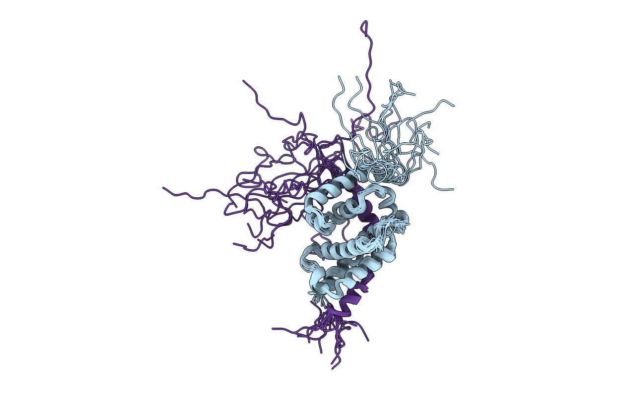
PDB ID:
2LFW
Keywords:
Title:
NMR structure of the PhyRSL-NepR complex from Sphingomonas sp. Fr1
Biological Source:
Source Organism(s):
Sphingomonas sp. Fr1 (Taxon ID: 907061)
Expression System(s):
Method Details:
Experimental Method:
Conformers Calculated:
20
Conformers Submitted:
15
Selection Criteria:
structures with the least restraint violations
