Deposition Date
1989-03-27
Release Date
1989-07-12
Last Version Date
2024-12-25
Entry Detail
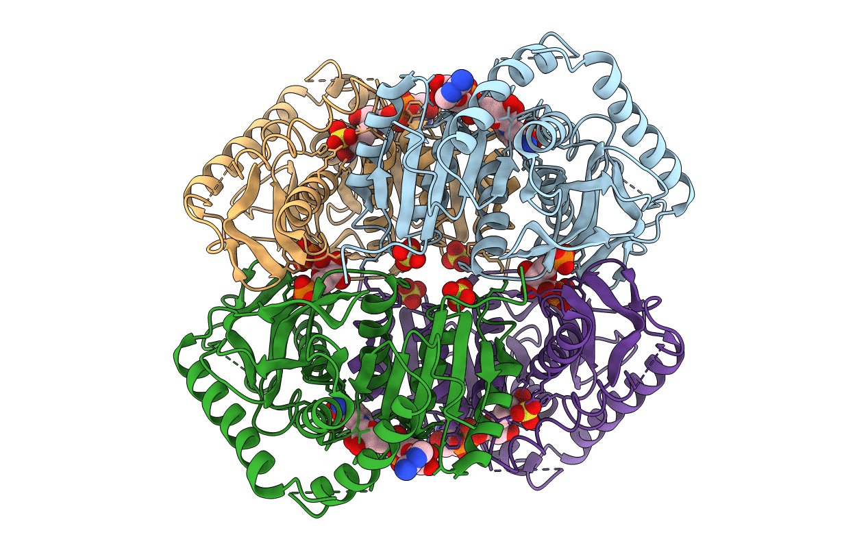
PDB ID:
2LDB
Keywords:
Title:
STRUCTURE DETERMINATION AND REFINEMENT OF BACILLUS STEAROTHERMOPHILUS LACTATE DEHYDROGENASE
Biological Source:
Source Organism(s):
Geobacillus stearothermophilus (Taxon ID: 1422)
Method Details: