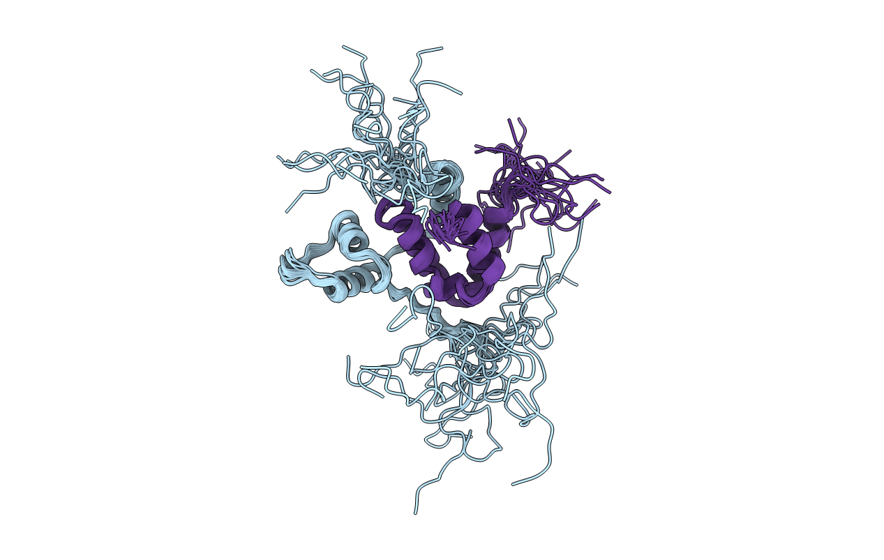
2LD7 image
Deposition Date 2011-05-16
Release Date 2011-06-15
Last Version Date 2024-05-15
Entry Detail
PDB ID:
2LD7
Keywords:
Title:
Solution structure of the mSin3A PAH3-SAP30 SID complex
Biological Source:
Source Organism:
Mus musculus (Taxon ID: 10090)
Host Organism:
Method Details:
Experimental Method:
Conformers Calculated:
80
Conformers Submitted:
20
Selection Criteria:
structures with the lowest energy
Macromolecular Entities
Polymer Type:polypeptide(L)
Molecule:Histone deacetylase complex subunit SAP30
Gene (Uniprot):Sap30
Chain IDs:A
Chain Length:94
Number of Molecules:1
Biological Source:Mus musculus
Polymer Type:polypeptide(L)
Molecule:Paired amphipathic helix protein Sin3a
Gene (Uniprot):Sin3a
Chain IDs:B
Chain Length:75
Number of Molecules:1
Biological Source:Mus musculus
Ligand Molecules
Primary Citation
Structure of the 30-kDa Sin3-associated protein (SAP30) in complex with the mammalian Sin3A corepressor and its role in nucleic acid binding.
J.Biol.Chem. 286 27814 27824 (2011)
PMID: 21676866 DOI: 10.1074/jbc.M111.252494

Abstact

The ∼2-megadalton evolutionarily conserved histone deacetylase-associated Rpd3L/Sin3L complex plays critical roles in altering the histone code and repressing transcription of a broad range of genes involved in many aspects of cellular physiology. Targeting of this complex to specific regions of the genome is presumed to rely on interactions involving one or more of at least 10 distinct subunits in the complex. Here we describe the solution structure of the complex formed by the interacting domains of two constitutively associated subunits, mSin3A and SAP30. The mSin3A paired amphipathic helix 3 (PAH3) domain in the complex adopts the left-handed four-helix bundle structure characteristic of PAH domains. The SAP30 Sin3 interaction domain (SID) binds to PAH3 via a tripartite structural motif, including a C-terminal helix that targets the canonical PAH hydrophobic cleft while two other helices and an N-terminal extension target a discrete surface formed largely by the PAH3 α2, α3, and α3' helices. The protein-protein interface is extensive (∼1400 Å(2)), accounting for the high affinity of the interaction and the constitutive association of the SAP30 subunit with the Rpd3L/Sin3L complex. We further show using NMR that the mSin3A PAH3-SAP30 SID complex can bind to nucleic acids, hinting at a role for a nucleolar localization sequence in the SID αA helix in targeting the Rpd3L/Sin3L complex for silencing ribosomal RNA genes.

Legend

Protein

Chemical

Disease

Primary Citation of related structures