Deposition Date
2011-05-12
Release Date
2011-06-22
Last Version Date
2024-10-30
Entry Detail
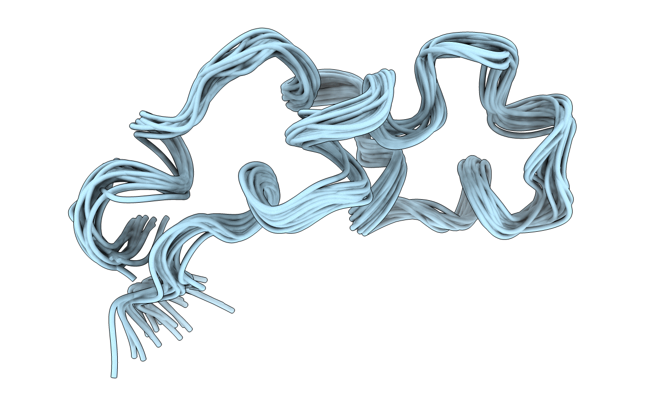
PDB ID:
2LCZ
Keywords:
Title:
NMR Structure of the Complete Internal Fusion Loop from Ebolavirus GP2 at pH 7.0
Biological Source:
Source Organism(s):
Zaire ebolavirus (Taxon ID: 186538)
Expression System(s):
Method Details:
Experimental Method:
Conformers Calculated:
200
Conformers Submitted:
20
Selection Criteria:
20 structures for lowest energy