2LCV image
Deposition Date 2011-05-10
Release Date 2011-06-29
Last Version Date 2024-05-15
Entry Detail
PDB ID:
2LCV
Title:
Structure of the Cytidine Repressor DNA-Binding Domain; an alternate calculation
Biological Source:
Source Organism:
Host Organism:
Method Details:
Experimental Method:
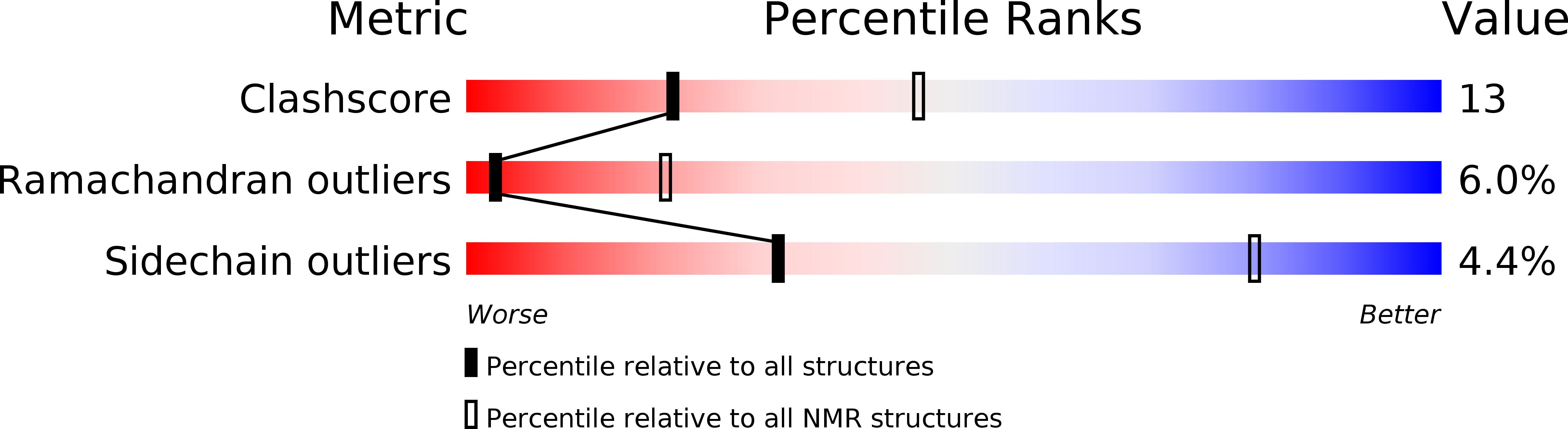
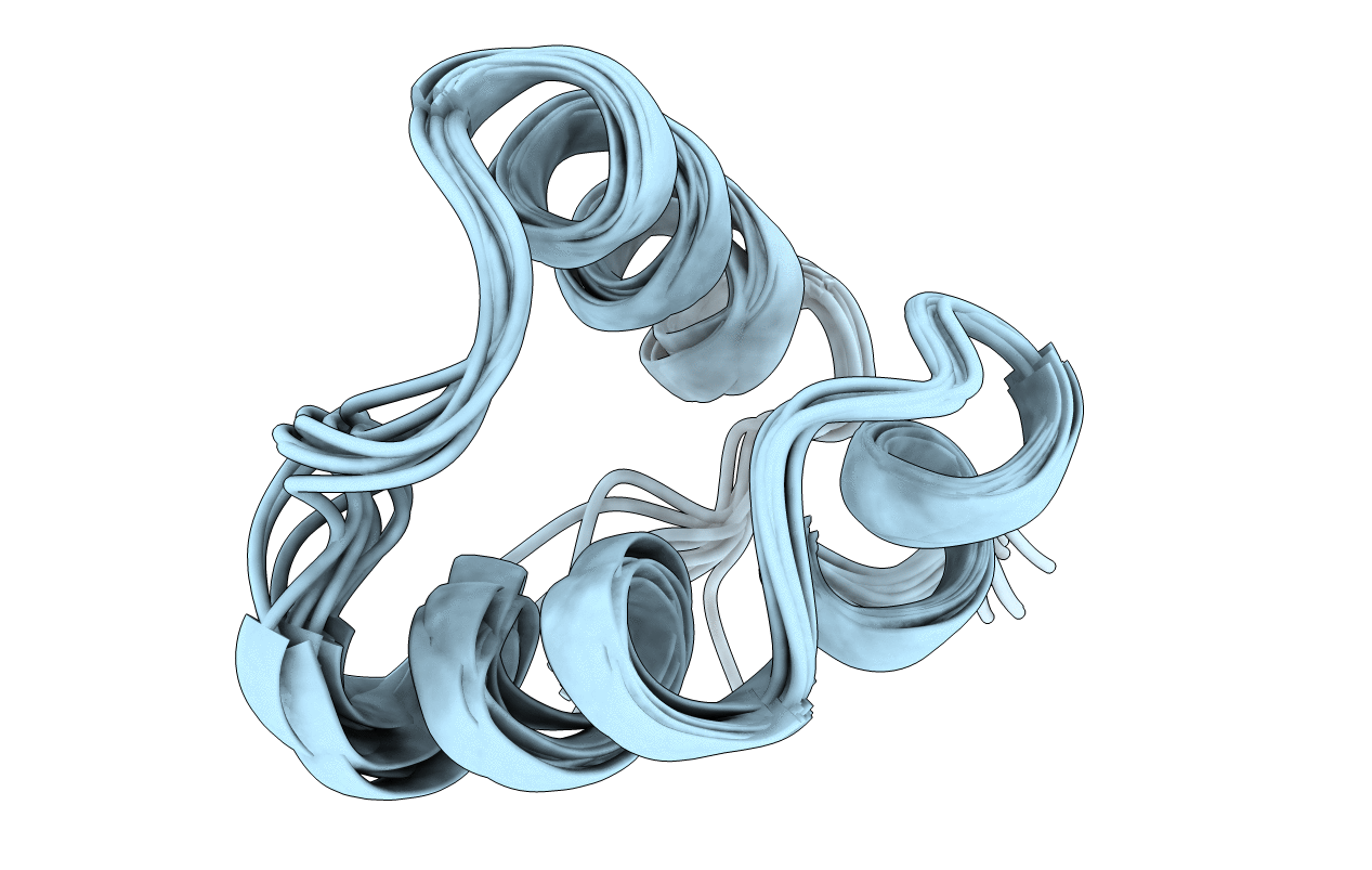
Conformers Calculated:
100
Conformers Submitted:
11
Selection Criteria:
structures with the lowest energy
Macromolecular Entities
Polymer Type:polypeptide(L)
Molecule:HTH-type transcriptional repressor CytR
Gene (Uniprot):cytR
Chain IDs:A
Chain Length:67
Number of Molecules:1
Biological Source:Escherichia coli K-12
Ligand Molecules
Primary Citation
Multiple Conformations of the Cytidine Repressor DNA-Binding Domain Coalesce to One upon Recognition of a Specific DNA Surface.
Biochemistry 50 6622 6632 (2011)
PMID: 21688840 DOI: 10.1021/bi200205v

Abstact

The cytidine repressor (CytR) is a member of the LacR family of bacterial repressors with distinct functional features. The Escherichia coli CytR regulon comprises nine operons whose palindromic operators vary in both sequence and, most significantly, spacing between the recognition half-sites. This suggests a strong likelihood that protein folding would be coupled to DNA binding as a mechanism to accommodate the variety of different operator architectures to which CytR is targeted. Such coupling is a common feature of sequence-specific DNA-binding proteins, including the LacR family repressors; however, there are no significant structural rearrangements upon DNA binding within the three-helix DNA-binding domains (DBDs) studied to date. We used nuclear magnetic resonance (NMR) spectroscopy to characterize the CytR DBD free in solution and to determine the high-resolution structure of a CytR DBD monomer bound specifically to one DNA half-site of the uridine phosphorylase (udp) operator. We find that the free DBD populates multiple distinct conformations distinguished by up to four sets of NMR peaks per residue. This structural heterogeneity is previously unknown in the LacR family. These stable structures coalesce into a single, more stable udp-bound form that features a three-helix bundle containing a canonical helix-turn-helix motif. However, this structure differs from all other LacR family members whose structures are known with regard to the packing of the helices and consequently their relative orientations. Aspects of CytR activity are unique among repressors; we identify here structural properties that are also distinct and that might underlie the different functional properties.

Legend

Protein

Chemical

Disease

Primary Citation of related structures