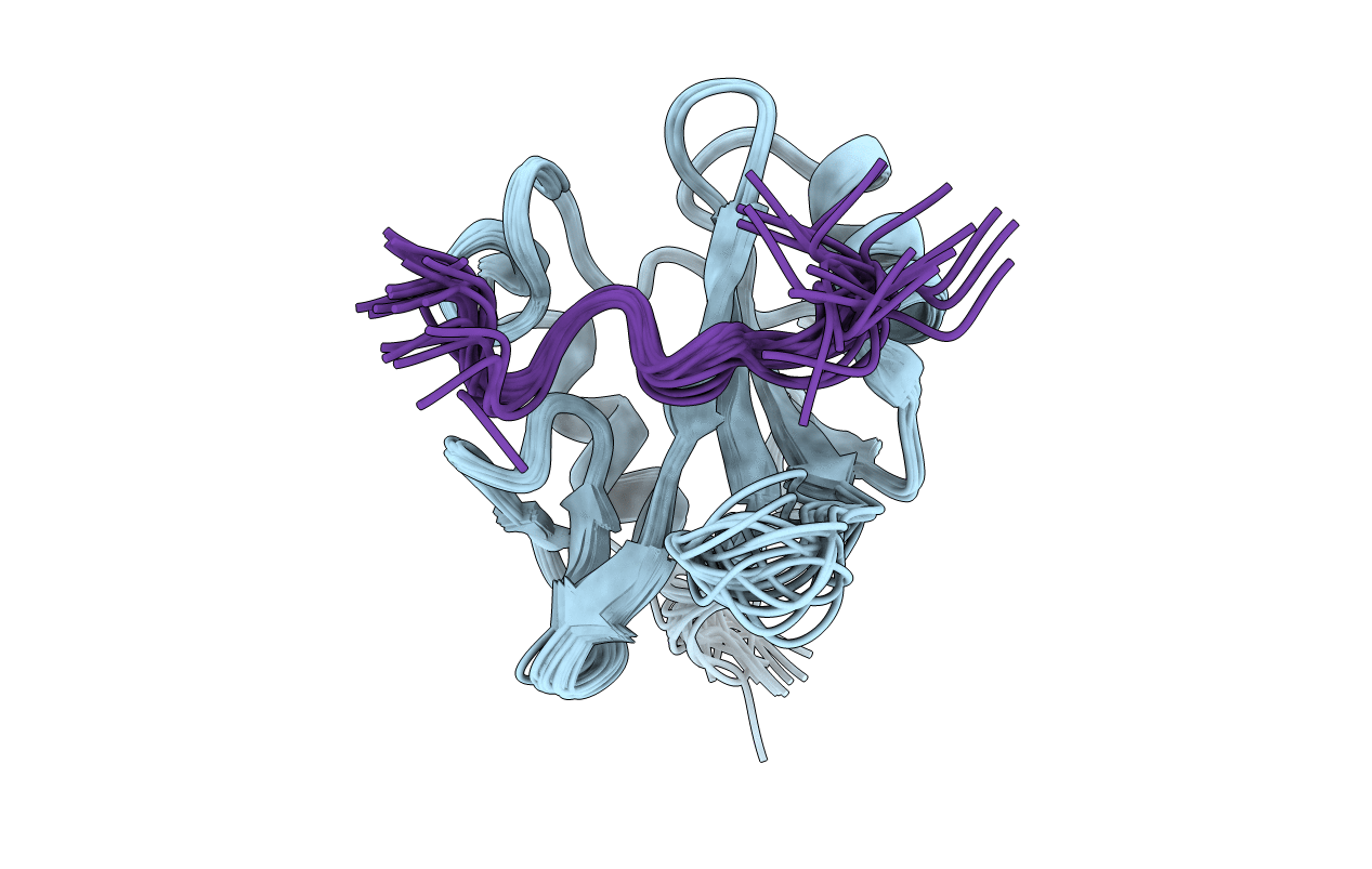
2LCT image
Deposition Date 2011-05-09
Release Date 2011-06-01
Last Version Date 2024-11-06
Entry Detail
PDB ID:
2LCT
Title:
Solution structure of the Vav1 SH2 domain complexed with a Syk-derived doubly phosphorylated peptide
Biological Source:
Source Organism:
Homo sapiens (Taxon ID: 9606)
Mus musculus (Taxon ID: 10090)
Host Organism:
Method Details:
Experimental Method:
Conformers Calculated:
100
Conformers Submitted:
20
Selection Criteria:
structures with the least restraint violations
Macromolecular Entities
Polymer Type:polypeptide(L)
Molecule:Proto-oncogene vav
Gene (Uniprot):VAV1
Chain IDs:A
Chain Length:107
Number of Molecules:1
Biological Source:Homo sapiens
Polymer Type:polypeptide(L)
Molecule:Tyrosine-protein kinase SYK
Gene (Uniprot):Syk
Chain IDs:B
Chain Length:13
Number of Molecules:1
Biological Source:Mus musculus
Modified Residue
Compound ID Chain ID Parent Comp ID Details 2D Image
PTR B TYR O-PHOSPHOTYROSINE
Ligand Molecules
Primary Citation
Two closely spaced tyrosines regulate NFAT signaling in B cells via Syk association with Vav.
Mol.Cell.Biol. 31 2984 2996 (2011)
PMID: 21606197 DOI: 10.1128/MCB.05043-11

Abstact

Activated Syk, an essential tyrosine kinase in B cell signaling, interacts with Vav guanine nucleotide exchange factors and regulates Vav activity through tyrosine phosphorylation. The Vav SH2 domain binds Syk linker B by an unusual recognition of two closely spaced Syk tyrosines: Y342 and Y346. The binding affinity is highest when both Y342 and Y346 are phosphorylated. An investigation in B cells of the dependence of Vav phosphorylation and NFAT activation on phosphorylation of Y342 and Y346 finds that cellular response levels match the relative binding affinities of the Vav1 SH2 domain for singly and doubly phosphorylated linker B peptides. This key result suggests that the uncommon recognition determinant of these two closely spaced tyrosines is a limiting factor in signaling. Interestingly, differences in affinities for binding singly and doubly phosphorylated peptides are reflected in the on rate, not the off rate. Such a control mechanism would be highly effective for regulating binding among competing Syk binding partners. The nuclear magnetic resonance (NMR) structure of Vav1 SH2 in complex with a doubly phosphorylated linker B peptide reveals diverse conformations associated with the unusual SH2 recognition of two phosphotyrosines. NMR relaxation indicates compensatory changes in loop fluctuations upon binding, with implications for nonphosphotyrosine interactions of Vav1 SH2.

Legend

Protein

Chemical

Disease

Primary Citation of related structures