2LC7 image
Deposition Date 2011-04-25
Release Date 2011-11-30
Last Version Date 2024-05-15
Entry Detail
PDB ID:
2LC7
Keywords:
Title:
Solution structure of the isolated Par-6 PDZ domain
Biological Source:
Source Organism:
Host Organism:
Method Details:
Experimental Method:
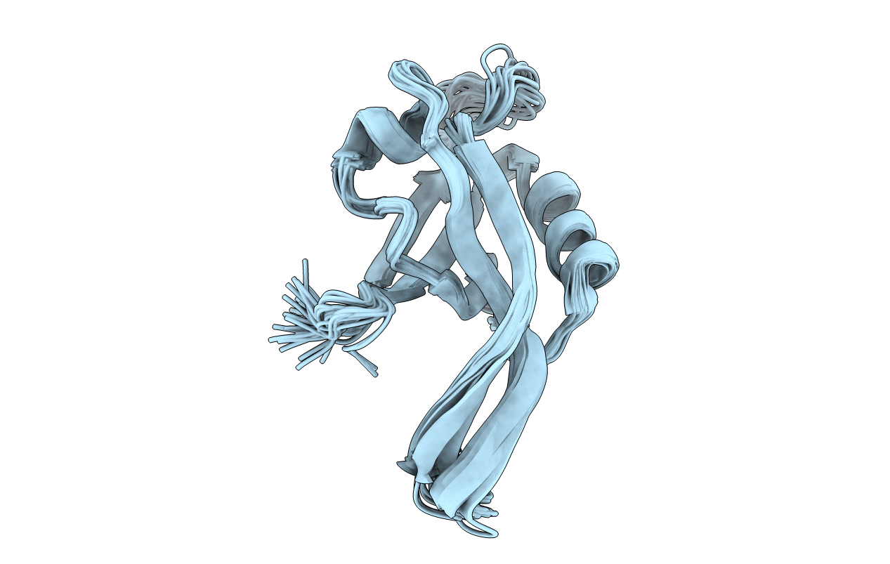
Conformers Calculated:
100
Conformers Submitted:
20
Selection Criteria:
target function
Macromolecular Entities
Structures with similar UniProt ID
Protein Blast
Polymer Type:polypeptide(L)
Molecule:Par-6
Gene (Uniprot):par-6
Chain IDs:A
Chain Length:102
Number of Molecules:1
Biological Source:Drosophila melanogaster
Ligand Molecules
Primary Citation
A Conformational Switch in the CRIB-PDZ Module of Par-6.
Structure 19 1711 1722 (2011)
PMID: 22078569 DOI: 10.1016/j.str.2011.07.018

Abstact

Here, we report a novel mechanism of PDZ (PSD-95/Dlg/ZO-1) domain regulation that distorts a conserved element of PDZ ligand recognition. The polarity regulator Par-6 assembles a conserved multiprotein complex and is directly modulated by the Rho GTPase Cdc42. Cdc42 binds the adjacent Cdc42/Rac interactive binding (CRIB) and PDZ domains of Par-6, increasing C-terminal ligand binding affinity by 10-fold. By solving structures of the isolated PDZ domain and a disulfide-stabilized CRIB-PDZ, we detected a conformational switch that controls affinity by altering the configuration of the conserved "GLGF" loop. As a result, lysine 165 is displaced from the PDZ core by an adjacent hydrophobic residue, disrupting coordination of the PDZ ligand-binding cleft. Stabilization of the CRIB:PDZ interface restores K165 to its canonical location in the binding pocket. We conclude that a unique "dipeptide switch" in the Par-6 PDZ transmits a signal for allosteric activation to the ligand-binding pocket.

Legend

Protein

Chemical

Disease

Primary Citation of related structures
Feedback Form
Name
Email
Institute
Feedback