2LC5 image
Deposition Date 2011-04-23
Release Date 2011-06-08
Last Version Date 2024-05-01
Entry Detail
PDB ID:
2LC5
Title:
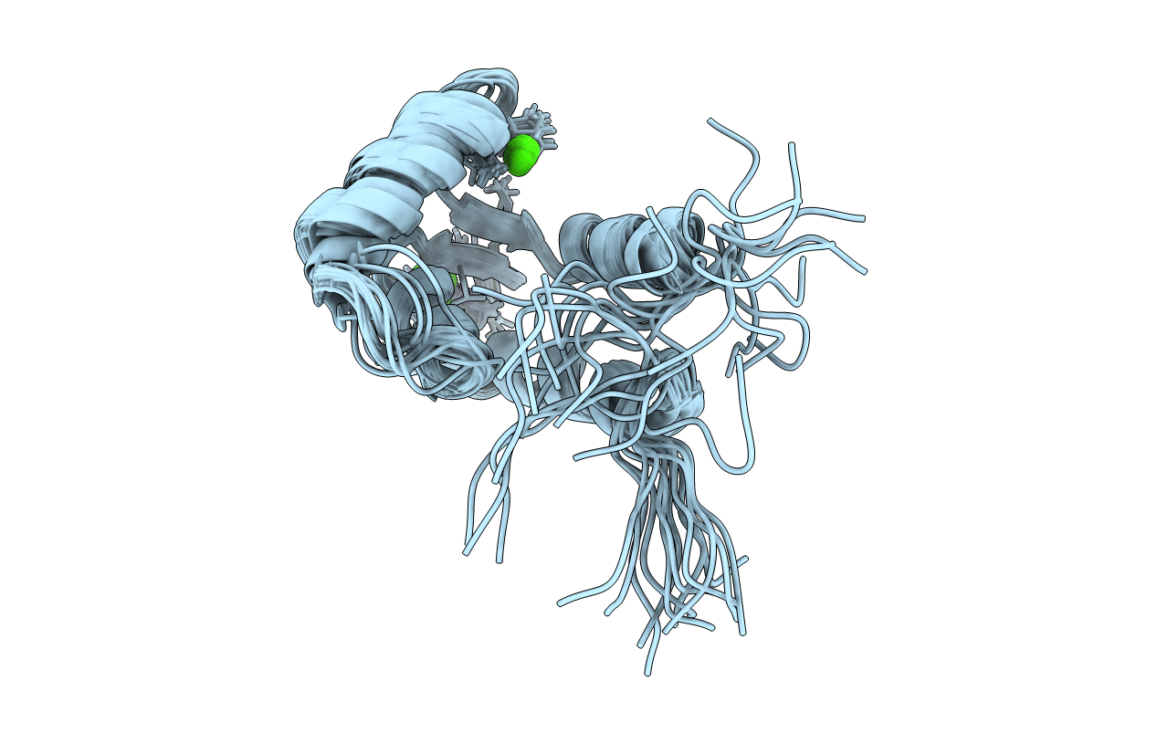
Calmodulin-like Protein from Entamoeba histolytica: Solution Structure and Calcium-Binding Properties of a Partially Folded Protein
Biological Source:
Source Organism:
Host Organism:
Method Details:
Experimental Method:
Conformers Calculated:
100
Conformers Submitted:
20
Selection Criteria:
STRUCTURES WITH THE LOWEST ENER
Macromolecular Entities
Polymer Type:polypeptide(L)
Molecule:Calmodulin, putative
Gene (Uniprot):EHI_100270
Chain IDs:A
Chain Length:151
Number of Molecules:1
Biological Source:Entamoeba histolytica
Ligand Molecules
Primary Citation
Calmodulin like protein from Entamoeba histolytica: solution structure and calcium binding properties of a partially folded protein.
Biochemistry 50 181 193 (2010)
PMID: 21114322 DOI: 10.1021/bi101411q

Abstact

The mechanism of Ca(2+)-signaling in the protozoan parasite Entamoeba histolytica is yet to be understood as many of the key regulators are still to be identified. E. histolytica encodes a number of multi-EF-hand Ca(2+)-binding proteins (EhCaBPs). Functionally only one of these molecules, EhCaBP1, has been characterized to date. The calmodulin-like protein from E. histolytica (abbreviated as EhCaM or EhCaBP3) is a 17.23 kDa monomeric protein that shows maximum sequence identity with heterologous calmodulins (CaMs). Though CaM activity has been biochemically shown in E. histolytica, there are no reports on the presence of a typical CaM. In an attempt to understand the structural and functional similarity of EhCaM with CaM, we have determined the three-dimensional (3D) solution structure of EhCaM using NMR. The EhCaM has a well-folded N-terminal domain and an unstructured C-terminal counterpart. Further, it sequentially binds only two calcium ions, an unusual mode of Ca(2+)-binding among the known CaBPs, notably both in the N-terminal domain of EhCaM. Further, EhCaM is present in the nucleus in addition to the cytoplasm as detected by immunofluorescence staining, unlike other EhCaBPs that are detected only in the cytoplasm. Therefore, this protein is likely to have a different function. The presence of unusual and a diverse set of CaBPs in E. histolytica suggests a distinct Ca(2+)-signaling process in E. histolytica. The results reported here help in understanding the structure-function relationship of CaBPs including their Ca(2+)-binding properties.

Legend

Protein

Chemical

Disease

Primary Citation of related structures