2LBF image
Deposition Date 2011-03-30
Release Date 2011-12-14
Last Version Date 2024-05-29
Entry Detail
PDB ID:
2LBF
Title:
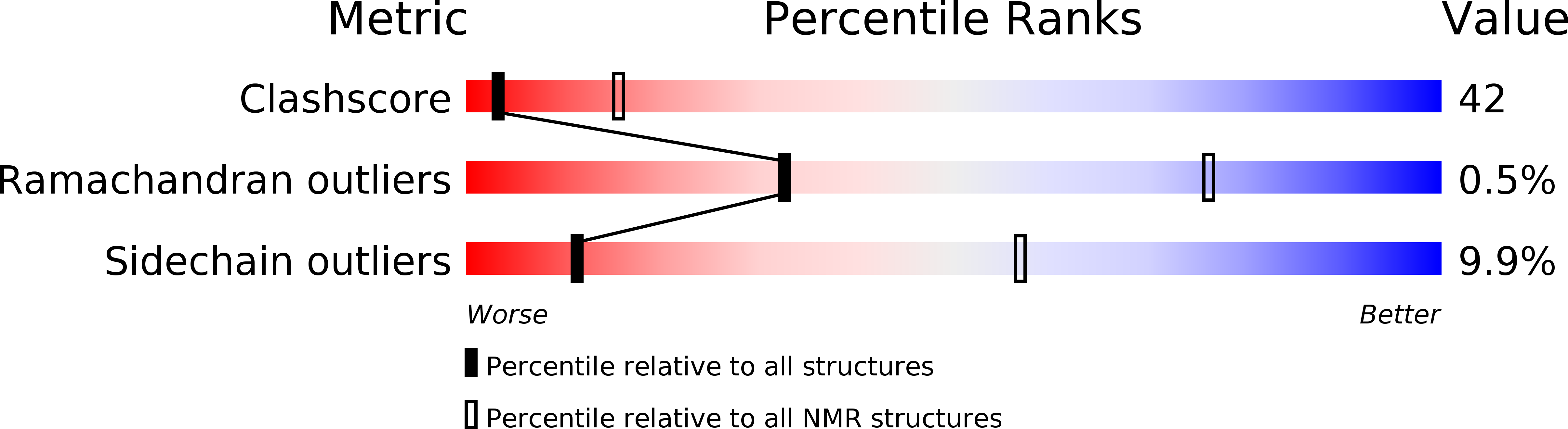
Solution structure of the dimerization domain of human ribosomal protein P1/P2 heterodimer
Biological Source:
Source Organism:
Homo sapiens (Taxon ID: 9606)
Host Organism:
Method Details:
Experimental Method:
Conformers Calculated:
100
Conformers Submitted:
10
Selection Criteria:
structures with the least restraint violations
Macromolecular Entities
Polymer Type:polypeptide(L)
Molecule:60S acidic ribosomal protein P1
Gene (Uniprot):RPLP1
Chain IDs:A
Chain Length:69
Number of Molecules:1
Biological Source:Homo sapiens
Polymer Type:polypeptide(L)
Molecule:60S acidic ribosomal protein P2
Gene (Uniprot):RPLP2
Chain IDs:B
Chain Length:70
Number of Molecules:1
Biological Source:Homo sapiens
Ligand Molecules
Primary Citation
Solution structure of the dimerization domain of the eukaryotic stalk P1/P2 complex reveals the structural organization of eukaryotic stalk complex
Nucleic Acids Res. ? ? ? (2011)
PMID: 22135285 DOI: 10.1093/nar/gkr1143

Abstact

The lateral ribosomal stalk is responsible for the kingdom-specific binding of translation factors and activation of GTP hydrolysis during protein synthesis. The eukaryotic stalk is composed of three acidic ribosomal proteins P0, P1 and P2. P0 binds two copies of P1/P2 hetero-dimers to form a pentameric P-complex. The structure of the eukaryotic stalk is currently not known. To provide a better understanding on the structural organization of eukaryotic stalk, we have determined the solution structure of the N-terminal dimerization domain (NTD) of P1/P2 hetero-dimer. Helix-1, -2 and -4 from each of the NTD-P1 and NTD-P2 form the dimeric interface that buries 2200 A(2) of solvent accessible surface area. In contrast to the symmetric P2 homo-dimer, P1/P2 hetero-dimer is asymmetric. Three conserved hydrophobic residues on the surface of NTD-P1 are replaced by charged residues in NTD-P2. Moreover, NTD-P1 has an extra turn in helix-1, which forms extensive intermolecular interactions with helix-1 and -4 of NTD-P2. Truncation of this extra turn of P1 abolished the formation of P1/P2 hetero-dimer. Systematic truncation studies suggest that P0 contains two spine-helices that each binds one copy of P1/P2 hetero-dimer. Modeling studies suggest that a large hydrophobic cavity, which can accommodate the loop between the spine-helices of P0, can be found on NTD-P1 but not on NTD-P2 when the helix-4 adopts an 'open' conformation. Based on the asymmetric properties of NTD-P1/NTD-P2, a structural model of the eukaryotic P-complex with P2/P1:P1/P2 topology is proposed.

Legend

Protein

Chemical

Disease

Primary Citation of related structures