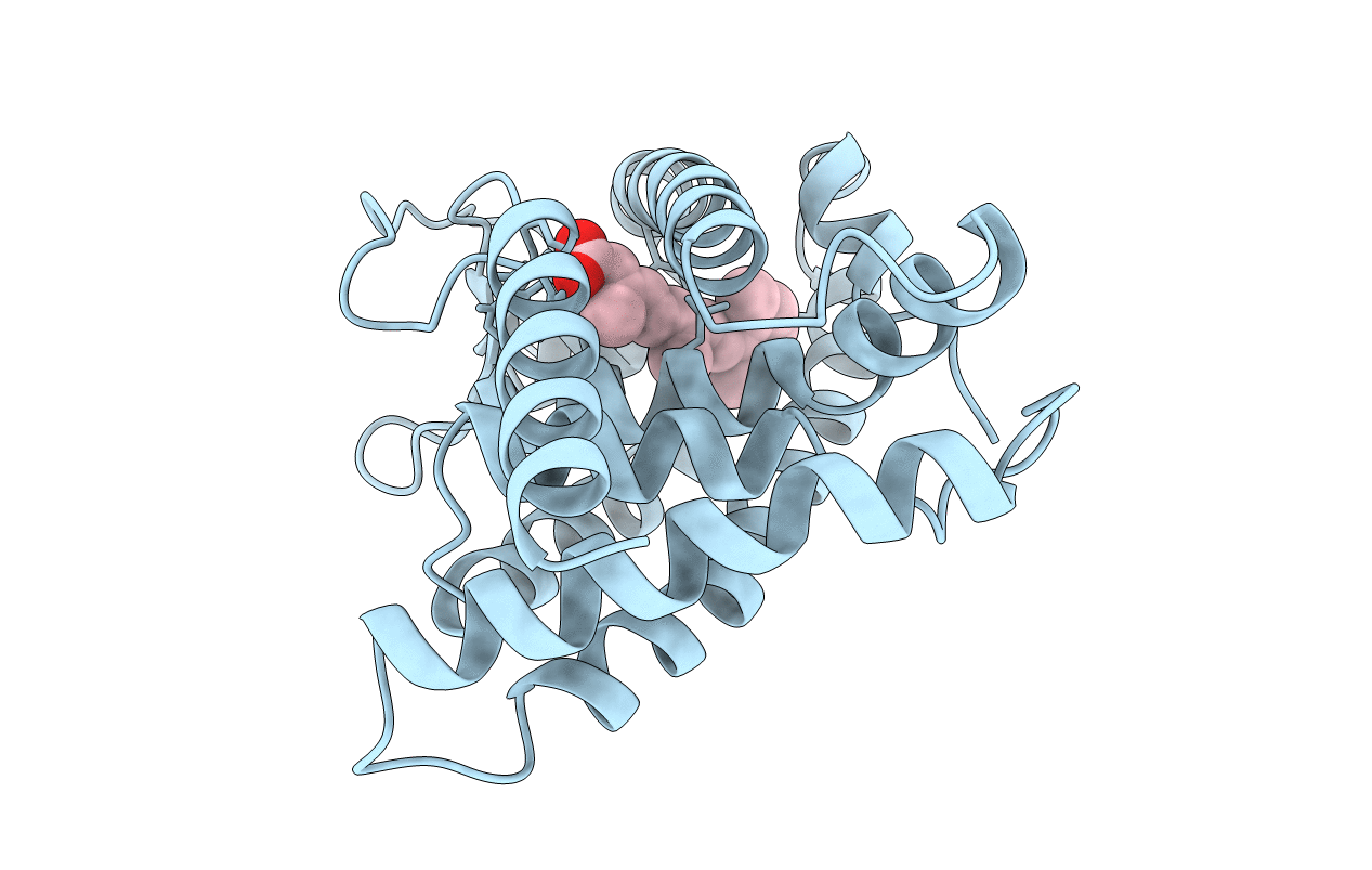
2LBD image
Entry Detail
PDB ID:
2LBD
Title:
LIGAND-BINDING DOMAIN OF THE HUMAN RETINOIC ACID RECEPTOR GAMMA BOUND TO ALL-TRANS RETINOIC ACID
Biological Source:
Source Organism:
PDB Version:
Deposition Date:
1997-08-19
Release Date:
1997-11-12
Method Details:
Experimental Method:
Resolution:
2.06 Å
R-Value Free:
0.31
R-Value Work:
0.21
R-Value Observed:
0.21
Space Group:
P 41 21 2
Macromolecular Entities
Polymer Type:polypeptide(L)
Description:RETINOIC ACID RECEPTOR GAMMA
Chain IDs:A
Chain Length:267
Number of Molecules:1
Biological Source:Homo sapiens
Ligand Molecules
Primary Citation
Crystal structure of the RAR-gamma ligand-binding domain bound to all-trans retinoic acid.
Nature 378 681 689 (1995)
PMID: 7501014 DOI: 10.1038/378681a0

Abstact

The 2.0-A crystal structure of the ligand-binding domain (LBD) of the human retinoic acid receptor (RAR)-gamma bound to all-trans retinoic acid reveals the ligand-binding interactions and suggests an electrostatic guidance mechanism. The overall fold is similar to that of the human RXR-alpha apo-LBD, except for the carboxy-terminal part which folds back towards the LBD core, contributing to the hydrophobic ligand pocket and 'sealing' its entry site. We propose a 'mouse trap' mechanism whereby a ligand-induced conformational transition repositions the amphipathic alpha-helix of the AF-2 activating domain and forms a transcriptionally active receptor.

Legend

Protein

Chemical

Disease

Primary Citation of related structures