Deposition Date
1993-06-10
Release Date
1993-10-31
Last Version Date
2024-02-21
Entry Detail
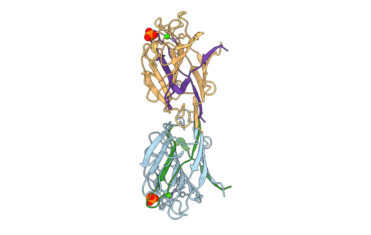
PDB ID:
2LAL
Keywords:
Title:
CRYSTAL STRUCTURE DETERMINATION AND REFINEMENT AT 2.3 ANGSTROMS RESOLUTION OF THE LENTIL LECTIN
Biological Source:
Source Organism(s):
Lens culinaris (Taxon ID: 3864)
Method Details:
Experimental Method:
Resolution:
1.80 Å
R-Value Observed:
0.18
Space Group:
P 21 21 21