
Deposition Date
2011-03-08
Release Date
2011-11-30
Last Version Date
2024-05-29
Entry Detail
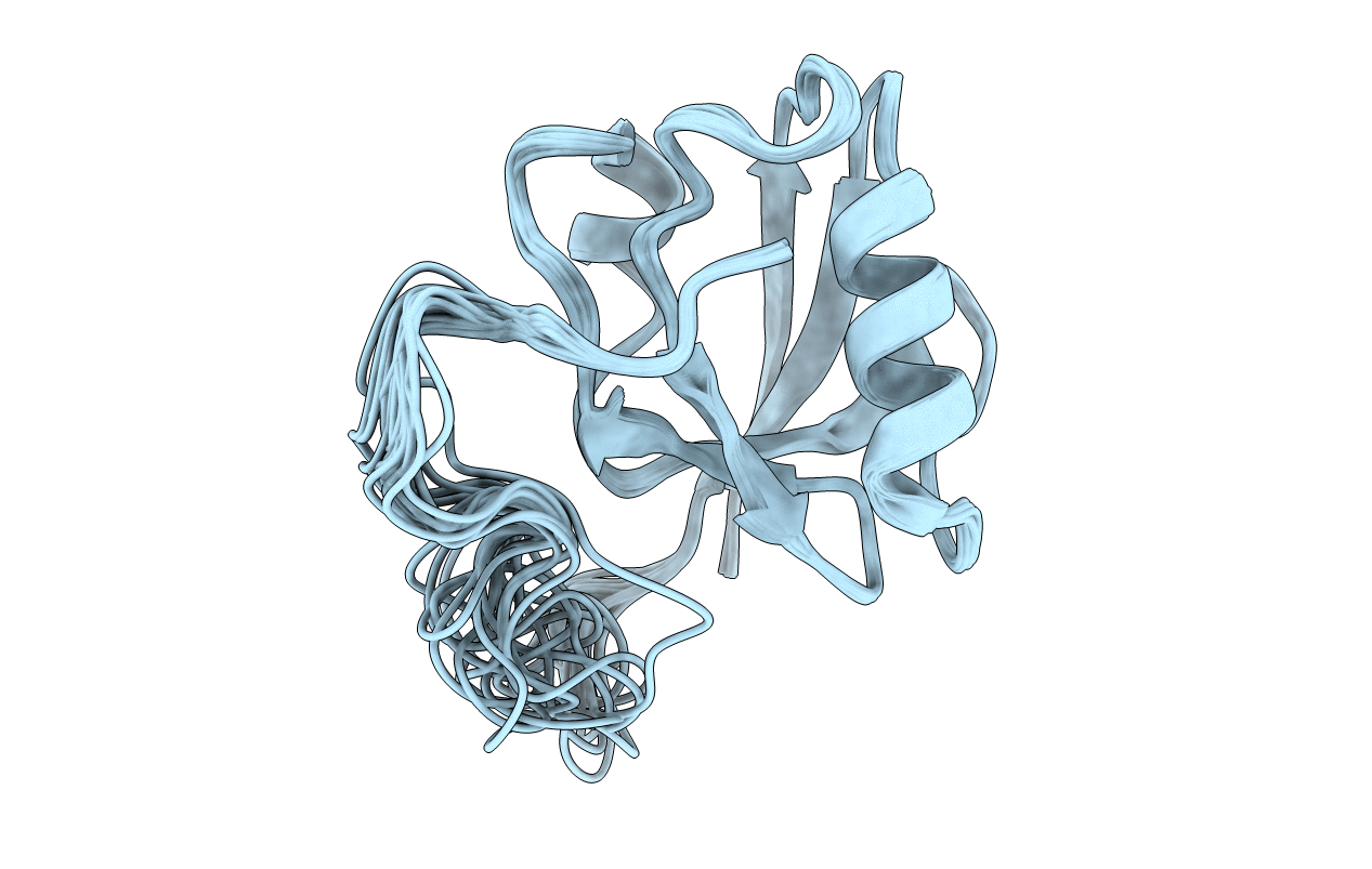
PDB ID:
2LA8
Keywords:
Title:
Solution structure of INAD PDZ5 complexed with Kon-tiki peptide
Biological Source:
Source Organism(s):
Drosophila melanogaster (Taxon ID: 7227)
Expression System(s):
Method Details:
Experimental Method:
Conformers Calculated:
200
Conformers Submitted:
20
Selection Criteria:
structures with the lowest energy
