
Deposition Date
2011-01-27
Release Date
2011-04-06
Last Version Date
2024-10-30
Entry Detail
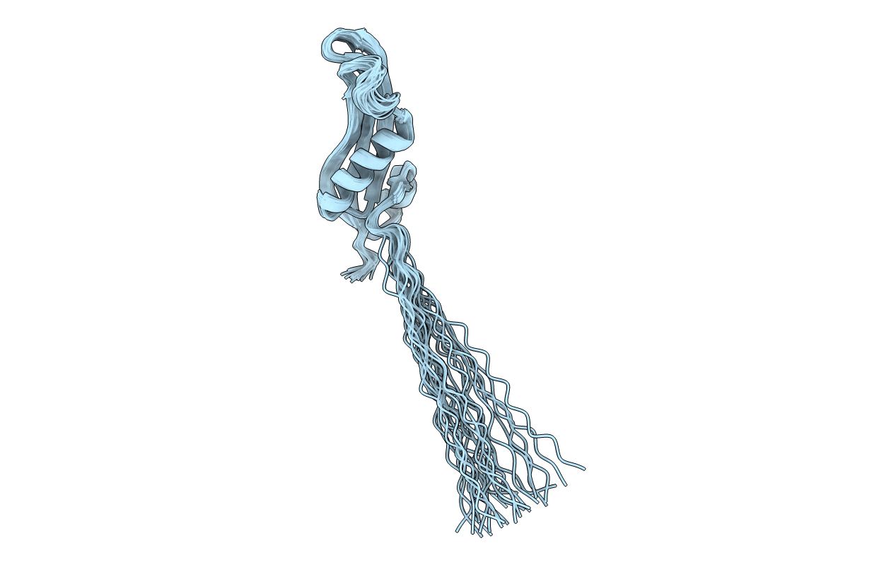
PDB ID:
2L8Y
Keywords:
Title:
Solution structure of the E. coli outer membrane protein RcsF (periplasmatic domain)
Biological Source:
Source Organism(s):
Escherichia coli (Taxon ID: 83333)
Expression System(s):
Method Details:
Experimental Method:
Conformers Calculated:
200
Conformers Submitted:
25
Selection Criteria:
structures with the lowest energy
