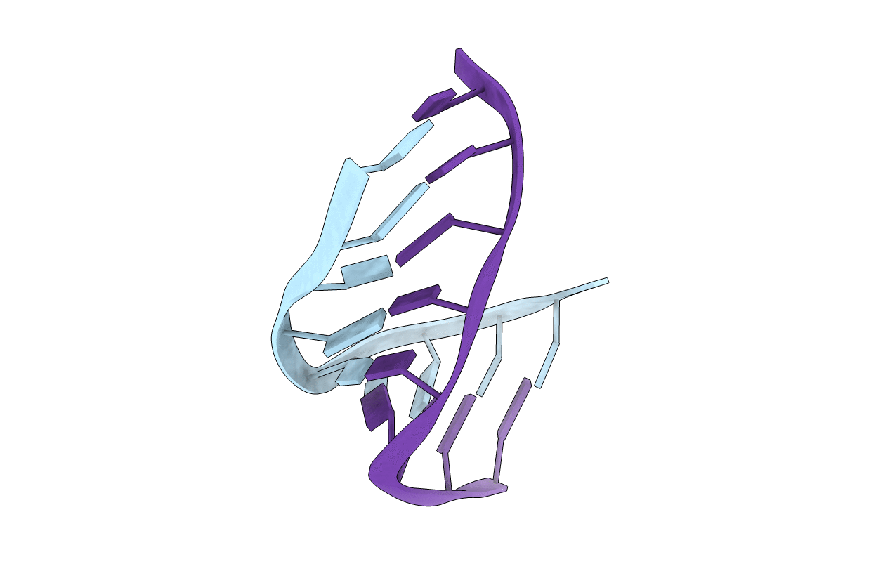
2L8C image
Deposition Date 2011-01-07
Release Date 2011-02-23
Last Version Date 2024-05-15
Entry Detail
PDB ID:
2L8C
Keywords:
Title:
NMR Spectroscopy and Molecular Dynamics Simulation of r(CCGCUGCGG)2 Reveal a Dynamic UU Internal Loop Found in Myotonic Dystrophy Type 1 - UU pair with zero hydrogen bond pairs
Biological Source:
Source Organism:
Method Details:
Experimental Method:
Conformers Calculated:
25
Conformers Submitted:
1
Selection Criteria:
structures with acceptable covalent geometry
Macromolecular Entities
Polymer Type:polyribonucleotide
Molecule:RNA (5'-R(*CP*CP*GP*CP*UP*GP*CP*GP*G)-3')
Chain IDs:A, B
Chain Length:9
Number of Molecules:2
Biological Source:synthetic construct
Ligand Molecules
Primary Citation
NMR Spectroscopy and Molecular Dynamics Simulation of r(CCGCUGCGG)(2) Reveal a Dynamic UU Internal Loop Found in Myotonic Dystrophy Type 1.
Biochemistry 50 599 601 (2011)
PMID: 21204525 DOI: 10.1021/bi101896j

Abstact

The NMR structure of an RNA with a copy of the 5'CUG/3'GUC motif found in the triplet repeating disorder myotonic dystrophy type 1 (DM1) is disclosed. The lowest energy conformation of the UU pair is a single-hydrogen bond structure; however, the UU protons undergo exchange indicating structural dynamics. Molecular dynamics simulations show that the single hydrogen bond structure is the most populated one but the UU pair interconverts among zero, one, and two hydrogen bond pairs. These studies have implications for the recognition of the DM1 RNA by small molecules and proteins.

Legend

Protein

Chemical

Disease

Primary Citation of related structures