2L7L image
Deposition Date 2010-12-13
Release Date 2011-05-18
Last Version Date 2024-05-15
Entry Detail
PDB ID:
2L7L
Title:
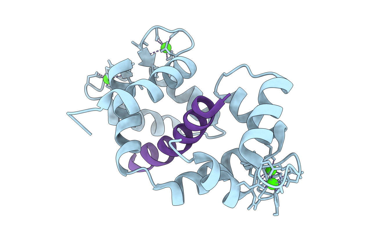
Solution structure of Ca2+/calmodulin complexed with a peptide representing the calmodulin-binding domain of calmodulin kinase I
Biological Source:
Source Organism:
Homo sapiens (Taxon ID: 9606)
Rattus norvegicus (Taxon ID: 10116)
Host Organism:
Method Details:
Experimental Method:
Conformers Calculated:
100
Conformers Submitted:
1
Selection Criteria:
structures with the lowest energy
Macromolecular Entities
Structures with similar UniProt ID
Protein Blast
Polymer Type:polypeptide(L)
Molecule:Calmodulin
Chain IDs:A
Chain Length:148
Number of Molecules:1
Biological Source:Homo sapiens
Structures with similar UniProt ID
Protein Blast
Polymer Type:polypeptide(L)
Molecule:Calcium/calmodulin-dependent protein kinase type 1
Gene (Uniprot):Camk1
Chain IDs:B
Chain Length:22
Number of Molecules:1
Biological Source:Rattus norvegicus
Ligand Molecules
Primary Citation
Fast methionine-based solution structure determination of calcium-calmodulin complexes.
J.Biomol.Nmr 50 71 81 (2011)
PMID: 21360154 DOI: 10.1007/s10858-011-9495-3

Abstact

Here we present a novel NMR method for the structure determination of calcium-calmodulin (Ca(2+)-CaM)-peptide complexes from a limited set of experimental restraints. A comparison of solved CaM-peptide structures reveals invariability in CaM's backbone conformation and a structural plasticity in CaM's domain orientation enabled by a flexible linker. Knowing this, the collection and analysis of an extensive set of NOESY spectra is redundant. Although RDCs can define CaM domain orientation in the complex, they lack the translational information required to position the domains on the bound peptide and highlight the necessity of intermolecular NOEs. Here we employ a specific isotope labeling strategy in which the role of methionine in CaM-peptide interactions is exploited to collect these critical NOEs. By (1)H, (13)C-labeling the methyl groups of deuterated methionine against a (2)H, (12)C background, we can acquire a (13)C-edited NOESY characterized by simplified, easily analyzable spectra. Together with measured CaM backbone H(N)-N RDCs and intrapeptide NOE-based distances, these intermolecular NOEs provide restraints for a low temperature torsion-angle dynamics and simulated annealing protocol used to calculate the complex structure. We have applied our method to a CaM complex previously solved through X-ray crystallography: Ca(2+)-CaM bound to the CaM kinase I peptide (PDB code: 1MXE). The resulting structure has a backbone RMSD of 1.6 Å to that previously published. We have also used this test complex to investigate the importance of homologous model selection on the calculated outcome. In addition to having application for fast complex structure determination, this method can be used to determine the structures of difficult complexes characterized by chemical shift overlap and broad signals for which the traditional method based on the use of fully (13)C, (15)N-labeled CaM fails.

Legend

Protein

Chemical

Disease

Primary Citation of related structures