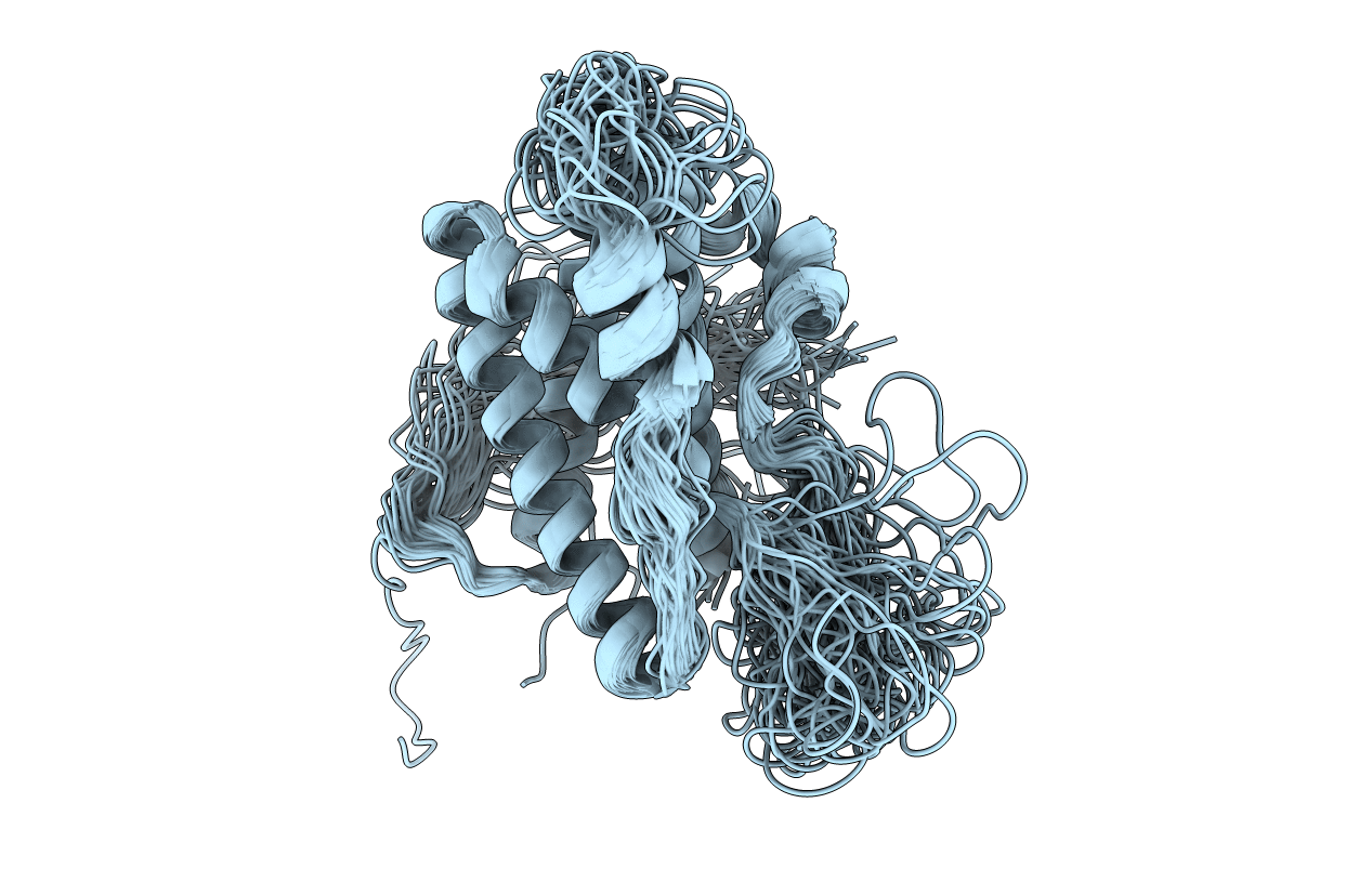
2L3Y image
Deposition Date 2010-09-25
Release Date 2011-09-28
Last Version Date 2024-10-30
Entry Detail
PDB ID:
2L3Y
Keywords:
Title:
Solution structure of mouse IL-6
Biological Source:
Source Organism:
Mus musculus (Taxon ID: 10090)
Host Organism:
Method Details:
Experimental Method:
Conformers Calculated:
100
Conformers Submitted:
52
Selection Criteria:
structures with the least restraint violations
Macromolecular Entities
Structures with similar UniProt ID
Protein Blast
Polymer Type:polypeptide(L)
Molecule:Interleukin-6
Gene (Uniprot):Il6
Chain IDs:A
Chain Length:190
Number of Molecules:1
Biological Source:Mus musculus
Ligand Molecules
Primary Citation
Conservation of functional sites on interleukin-6 and implications for evolution of signaling complex assembly and therapeutic intervention.
J.Biol.Chem. 287 40043 40050 (2012)
PMID: 23027872 DOI: 10.1074/jbc.M112.405597

Abstact

A number of secreted cytokines, such as interleukin-6 (IL-6), are attractive targets for the treatment of inflammatory diseases. We have determined the solution structure of mouse IL-6 to assess the functional significance of apparent differences in the receptor interaction sites (IL-6Rα and gp130) suggested by the fairly low degree of sequence similarity with human IL-6. Structure-based sequence alignment of mouse IL-6 and human IL-6 revealed surprising differences in the conservation of the two distinct gp130 binding sites (IIa and IIIa), which suggests a primacy for site III-mediated interactions in driving initial assembly of the IL-6/IL-6Rα/gp130 ternary complex. This is further supported by a series of direct binding experiments, which clearly demonstrate a high affinity IL-6/IL-6Rα-gp130 interaction via site III but only weak binding via site II. Collectively, our findings suggest a pathway for the evolution of the hexameric, IL-6/IL-6Rα/gp130 signaling complex and strategies for therapeutic targeting. We propose that the signaling complex originally involved specific interactions between IL-6 and IL-6Rα (site I) and between the D1 domain of gp130 and IL-6/IL-6Rα (site III), with the later inclusion of interactions between the D2 and D3 domains of gp130 and IL-6/IL-6Rα (site II) through serendipity. It seems likely that IL-6 signaling benefited from the evolution of a multipurpose, nonspecific protein interaction surface on gp130, now known as the cytokine binding homology region (site II contact surface), which fortuitously contributes to stabilization of the IL-6/IL-6Rα/gp130 signaling complex.

Legend

Protein

Chemical

Disease

Primary Citation of related structures