2L3O image
Deposition Date 2010-09-20
Release Date 2011-08-10
Last Version Date 2024-10-30
Entry Detail
PDB ID:
2L3O
Keywords:
Title:
Solution structure of murine interleukin 3
Biological Source:
Source Organism:
Mus musculus (Taxon ID: 10090)
Host Organism:
Method Details:
Experimental Method:
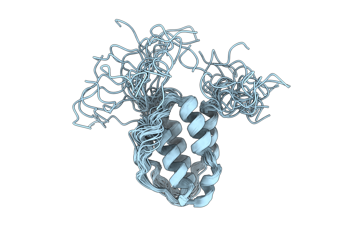
Conformers Calculated:
200
Conformers Submitted:
20
Selection Criteria:
on the basis of stereochemistry and energy considerations
Macromolecular Entities
Structures with similar UniProt ID
Protein Blast
Polymer Type:polypeptide(L)
Molecule:Interleukin 3
Mutagens:C105A
Chain IDs:A
Chain Length:127
Number of Molecules:1
Biological Source:Mus musculus
Ligand Molecules
Primary Citation
Murine interleukin-3: structure, dynamics, and conformational heterogeneity in solution.
Biochemistry 50 2464 2477 (2011)
PMID: 21329364 DOI: 10.1021/bi101810f

Abstact

Interleukin-3 (IL-3), a cytokine produced primarily by activated T-cells during immune responses, is a crucial regulator of allergic inflammation. The three-dimensional structure of murine IL-3 (mIL-3) has remained elusive owing to its poor solubility and strong tendency toward aggregation under solution conditions typically used for structural studies. Here we describe the solution properties and structure of mIL-3 determined by NMR using an engineered construct of mIL-3 (mIL-3(33-156)). mIL-3 adopts a four-helical bundle fold, typical of proteins belonging to the short-chain cytokine family, and features a core of highly conserved hydrophobic residues. While significant line broadening and peak disappearance were observed in NMR spectra at higher temperatures, there was no evidence for temperature-dependent changes of the oligomeric state of mIL-3(33-156). Further analysis of the temperature dependence of amide (1)H chemical shifts and backbone (15)N relaxation parameters, including (15)N relaxation dispersion, revealed the presence of significant conformational exchange and local conformational heterogeneity. Residues recently shown by mutagenesis to play key roles in β(IL-3) receptor recognition and activation, which are located within the α(A) and α(C) helices and aligned on one face of the mIL-3(33-156) structure, are relatively rigid. In contrast, pronounced conformational heterogeneity was observed for a cluster of residues located on the opposite side of mIL-3, which corresponds spatially to sites in the related cytokines human IL-3, IL-5, and GM-CSF that are known to mediate interactions with their respective α-receptor subunits. Such conformational heterogeneity may facilitate the interaction of mIL-3 with each of two naturally occurring mIL-3Rα isoforms, leading to structurally distinct high-affinity complexes.

Legend

Protein

Chemical

Disease

Primary Citation of related structures