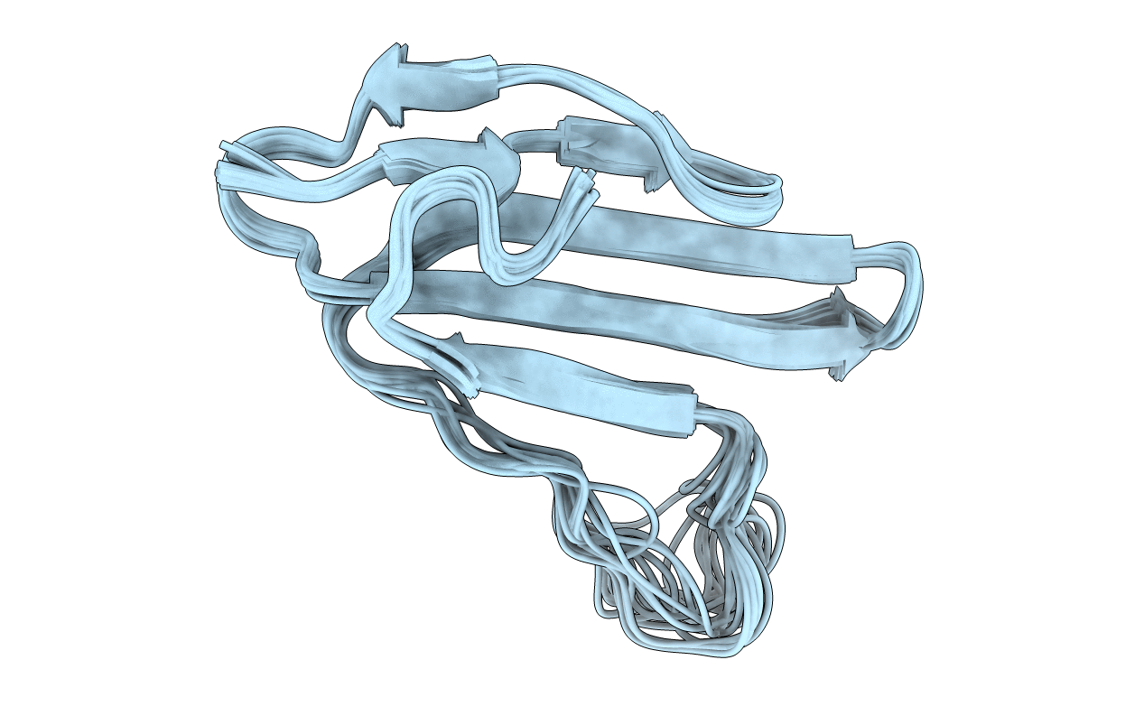
2L03 image
Deposition Date 2010-06-30
Release Date 2011-01-19
Last Version Date 2024-11-20
Entry Detail
PDB ID:
2L03
Keywords:
Title:
Spatial structure of water-soluble Lynx1
Biological Source:
Source Organism:
Homo sapiens (Taxon ID: 9606)
Host Organism:
Method Details:
Experimental Method:
Conformers Calculated:
200
Conformers Submitted:
20
Selection Criteria:
target function
Macromolecular Entities
Structures with similar UniProt ID
Protein Blast
Polymer Type:polypeptide(L)
Molecule:Ly-6/neurotoxin-like protein 1
Chain IDs:A
Chain Length:74
Number of Molecules:1
Biological Source:Homo sapiens
Ligand Molecules
Primary Citation
NMR structure and action on nicotinic acetylcholine receptors of water-soluble domain of human LYNX1
J.Biol.Chem. 286 10618 10627 (2011)
PMID: 21252236 DOI: 10.1074/jbc.M110.189100

Abstact

Discovery of proteins expressed in the central nervous system sharing the three-finger structure with snake α-neurotoxins provoked much interest to their role in brain functions. Prototoxin LYNX1, having homology both to Ly6 proteins and three-finger neurotoxins, is the first identified member of this family membrane-tethered by a GPI anchor, which considerably complicates in vitro studies. We report for the first time the NMR spatial structure for the water-soluble domain of human LYNX1 lacking a GPI anchor (ws-LYNX1) and its concentration-dependent activity on nicotinic acetylcholine receptors (nAChRs). At 5-30 μM, ws-LYNX1 competed with (125)I-α-bungarotoxin for binding to the acetylcholine-binding proteins (AChBPs) and to Torpedo nAChR. Exposure of Xenopus oocytes expressing α7 nAChRs to 1 μM ws-LYNX1 enhanced the response to acetylcholine, but no effect was detected on α4β2 and α3β2 nAChRs. Increasing ws-LYNX1 concentration to 10 μM caused a modest inhibition of these three nAChR subtypes. A common feature for ws-LYNX1 and LYNX1 is a decrease of nAChR sensitivity to high concentrations of acetylcholine. NMR and functional analysis both demonstrate that ws-LYNX1 is an appropriate model to shed light on the mechanism of LYNX1 action. Computer modeling, based on ws-LYNX1 NMR structure and AChBP x-ray structure, revealed a possible mode of ws-LYNX1 binding.

Legend

Protein

Chemical

Disease

Primary Citation of related structures