2KY1 image
Deposition Date 2010-05-13
Release Date 2010-06-02
Last Version Date 2024-05-22
Entry Detail
PDB ID:
2KY1
Keywords:
Title:
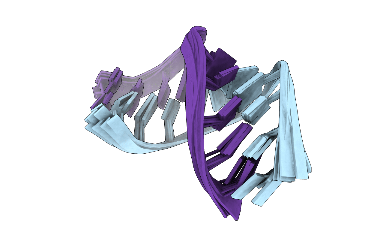
The Structure of RNA Internal Loops with Tandem AG Pairs: 5'UAGA/3'AGAU
Biological Source:
Source Organism:
(Taxon ID: ) (Taxon ID: )
Method Details:
Experimental Method:
Conformers Calculated:
40
Conformers Submitted:
10
Selection Criteria:
structures with the least restraint violations
Macromolecular Entities
Polymer Type:polyribonucleotide
Molecule:5'-R(*GP*AP*CP*UP*AP*GP*AP*GP*UP*CP*A)-3'
Chain IDs:A, B
Chain Length:11
Number of Molecules:2
Biological Source:
Ligand Molecules
Primary Citation
RNA internal loops with tandem AG pairs: the structure of the 5'GAGU/3'UGAG loop can be dramatically different from others, including 5'AAGU/3'UGAA.
Biochemistry 49 5817 5827 (2010)
PMID: 20481618 DOI: 10.1021/bi100332r

Abstact

Thermodynamic stabilities of 2 x 2 nucleotide tandem AG internal loops in RNA range from -1.3 to +3.4 kcal/mol at 37 degrees C and are not predicted well with a hydrogen-bonding model. To provide structural information to facilitate development of more sophisticated models for the sequence dependence of stability, we report the NMR solution structures of five RNA duplexes: (rGACGAGCGUCA)(2), (rGACUAGAGUCA)(2), (rGACAAGUGUCA)(2), (rGGUAGGCCA)(2), and (rGACGAGUGUCA)(2). The structures of these duplexes are compared to that of the previously solved (rGGCAGGCC)(2) (Wu, M., SantaLucia, J., Jr., and Turner, D. H. (1997) Biochemistry 36, 4449-4460). For loops bounded by Watson-Crick pairs, the AG and Watson-Crick pairs are all head-to-head imino-paired (cis Watson-Crick/Watson-Crick). The structures suggest that the sequence-dependent stability may reflect non-hydrogen-bonding interactions. Of the two loops bounded by G-U pairs, only the 5'UAGG/3'GGAU loop adopts canonical UG wobble pairing (cis Watson-Crick/Watson-Crick), with AG pairs that are only weakly imino-paired. Strikingly, the 5'GAGU/3'UGAG loop has two distinct duplex conformations, the major of which has both guanosine residues (G4 and G6 in (rGACGAGUGUCA)(2)) in a syn glycosidic bond conformation and forming a sheared GG pair (G4-G6*, GG trans Watson-Crick/Hoogsteen), both uracils (U7 and U7*) flipped out of the helix, and an AA pair (A5-A5*) in a dynamic or stacked conformation. These structures provide benchmarks for computational investigations into interactions responsible for the unexpected differences in loop free energies and structure.

Legend

Protein

Chemical

Disease

Primary Citation of related structures