2KVA image
Entry Detail
PDB ID:
2KVA
Title:
SOLUTION STRUCTURE OF CI-MPR ligand-free domain 5
Biological Source:
Source Organism:
Host Organism:
PDB Version:
Deposition Date:
2010-03-10
Release Date:
2010-07-07
Method Details:
Experimental Method:
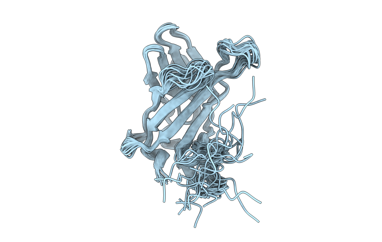
Conformers Calculated:
100
Conformers Submitted:
20
Selection Criteria:
target function
Macromolecular Entities
Polymer Type:polypeptide(L)
Description:Cation-independent mannose-6-phosphate receptor
Chain IDs:A
Chain Length:148
Number of Molecules:1
Biological Source:Bos taurus
Ligand Molecules
Primary Citation
Structural basis for recognition of phosphodiester-containing lysosomal enzymes by the cation-independent mannose 6-phosphate receptor.
Proc.Natl.Acad.Sci.USA 107 12493 12498 (2010)
PMID: 20615935 DOI: 10.1073/pnas.1004232107

Abstact

Mannose 6-phosphate (Man-6-P)-dependent trafficking is vital for normal development. The biogenesis of lysosomes, a major cellular site of protein, carbohydrate, and lipid catabolism, depends on the 300-kDa cation-independent Man-6-P receptor (CI-MPR) that transports newly synthesized acid hydrolases from the Golgi. The CI-MPR recognizes lysosomal enzymes bearing the Man-6-P modification, which arises by the addition of GlcNAc-1-phosphate to mannose residues and subsequent removal of GlcNAc by the uncovering enzyme (UCE). The CI-MPR also recognizes lysosomal enzymes that elude UCE maturation and instead display the Man-P-GlcNAc phosphodiester. This ability of the CI-MPR to target phosphodiester-containing enzymes ensures lysosomal delivery when UCE activity is deficient. The extracellular region of the CI-MPR is comprised of 15 repetitive domains and contains three distinct Man-6-P binding sites located in domains 3, 5, and 9, with only domain 5 exhibiting a marked preference for phosphodiester-containing lysosomal enzymes. To determine how the CI-MPR recognizes phosphodiesters, the structure of domain 5 was determined by NMR spectroscopy. Although domain 5 contains only three of the four disulfide bonds found in the other seven domains whose structures have been determined to date, it adopts the same fold consisting of a flattened beta-barrel. Structure determination of domain 5 bound to N-acetylglucosaminyl 6-phosphomethylmannoside, along with mutagenesis studies, revealed the residues involved in diester recognition, including Y679. These results show the mechanism by which the CI-MPR recognizes Man-P-GlcNAc-containing ligands and provides new avenues to investigate the role of phosphodiester-containing lysosomal enzymes in the biogenesis of lysosomes.

Legend

Protein

Chemical

Disease

Primary Citation of related structures