
Deposition Date
2010-02-04
Release Date
2010-04-07
Last Version Date
2024-10-30
Entry Detail
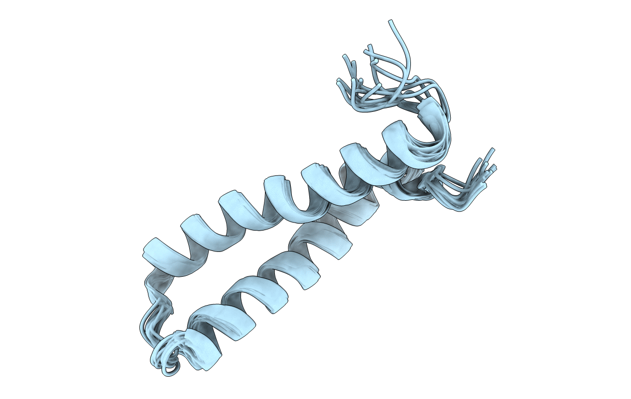
PDB ID:
2KTM
Keywords:
Title:
Solution NMR structure of H2H3 domain of ovine prion protein (residues 167-234)
Biological Source:
Source Organism(s):
Ovis aries (Taxon ID: 9940)
Expression System(s):
Method Details:
Experimental Method:
Conformers Calculated:
50
Conformers Submitted:
12
Selection Criteria:
structures with the lowest energy
