Deposition Date
2009-12-16
Release Date
2010-02-16
Last Version Date
2024-05-22
Entry Detail
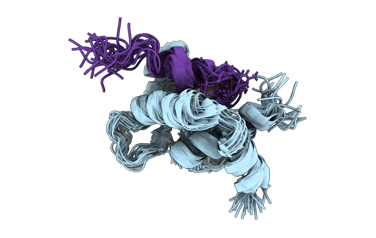
PDB ID:
2KRD
Keywords:
Title:
Solution Structure of the Regulatory Domain of Human Cardiac Troponin C in Complex with the Switch Region of cardiac Troponin I and W7
Biological Source:
Source Organism(s):
Homo sapiens (Taxon ID: 9606)
Expression System(s):
Method Details:
Experimental Method:
Conformers Calculated:
300
Conformers Submitted:
20
Selection Criteria:
structures with the lowest energy