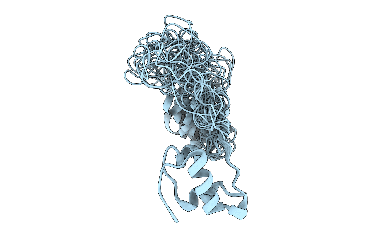
2KQP image
Entry Detail
PDB ID:
2KQP
Keywords:
Title:
NMR Structure of Proinsulin
Biological Source:
Source Organism:
Host Organism:
PDB Version:
Deposition Date:
2009-11-12
Release Date:
2010-01-26
Method Details:
Experimental Method:
Conformers Calculated:
90
Conformers Submitted:
20
Selection Criteria:
structures with acceptable covalent geometry
Macromolecular Entities
Polymer Type:polypeptide(L)
Description:Insulin
Mutations:H10D, P28K, K29P
Chain IDs:A
Chain Length:86
Number of Molecules:1
Biological Source:Homo sapiens
Ligand Molecules
Primary Citation
Solution structure of proinsulin: connecting domain flexibility and prohormone processing.
J.Biol.Chem. 285 7847 7851 (2010)
PMID: 20106974 DOI: 10.1074/jbc.C109.084921

Abstact

The folding of proinsulin, the single-chain precursor of insulin, ensures native disulfide pairing in pancreatic beta-cells. Mutations that impair folding cause neonatal diabetes mellitus. Although the classical structure of insulin is well established, proinsulin is refractory to crystallization. Here, we employ heteronuclear NMR spectroscopy to characterize a monomeric analogue. Proinsulin contains a native-like insulin moiety (A- and B-domains); the tethered connecting (C) domain (as probed by {(1)H}-(15)N nuclear Overhauser enhancements) is progressively less ordered. Although the BC junction is flexible, residues near the CA junction exhibit alpha-helical-like features. Relative to canonical alpha-helices, however, segmental (13)C(alpha/beta) chemical shifts are attenuated, suggesting that this junction and contiguous A-chain residues are molten. We propose that flexibility at each C-domain junction facilitates prohormone processing. Studies of protease SPC3 (PC1/3) suggest that C-domain sequences contribute to cleavage site selection. The structure of proinsulin provides a foundation for studies of insulin biosynthesis and its impairment in monogenic forms of diabetes mellitus.

Legend

Protein

Chemical

Disease

Primary Citation of related structures