Deposition Date
2009-10-28
Release Date
2010-05-12
Last Version Date
2024-05-08
Entry Detail
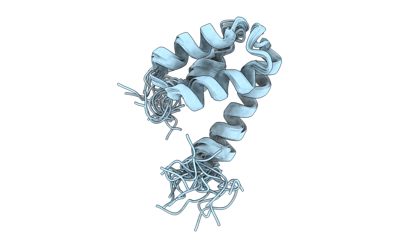
PDB ID:
2KQ6
Keywords:
Title:
The structure of the EF-hand domain of polycystin-2 suggests a mechanism for Ca2+-dependent regulation of polycystin-2 channel activity
Biological Source:
Source Organism(s):
Homo sapiens (Taxon ID: 9606)
Expression System(s):
Method Details:
Experimental Method:
Conformers Calculated:
80
Conformers Submitted:
20
Selection Criteria:
structures with the lowest energy and least restraint violations